The olfactory nerve has a role in the body temperature and brain cytokine responses to influenza virus
- PMID: 19836444
- PMCID: PMC2818451
- DOI: 10.1016/j.bbi.2009.10.007
The olfactory nerve has a role in the body temperature and brain cytokine responses to influenza virus
Abstract
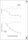
Mouse-adapted human influenza virus is detectable in the olfactory bulbs of mice within hours after intranasal challenge and is associated with enhanced local cytokine mRNA and protein levels. To determine whether signals from the olfactory nerve influence the unfolding of the acute phase response (APR), we surgically transected the olfactory nerve in mice prior to influenza infection. We then compared the responses of olfactory-nerve-transected (ONT) mice to those recorded in sham-operated control mice using measurements of body temperature, food intake, body weight, locomotor activity and immunohistochemistry for cytokines and the viral antigen, H1N1. ONT did not change baseline body temperature (Tb); however, the onset of virus-induced hypothermia was delayed for about 13 h in the ONT mice. Locomotor activity, food intake and body weights of the two groups were similar. At 15 h post-challenge fewer viral antigen-immunoreactive (IR) cells were observed in the olfactory bulb (OB) of ONT mice compared to sham controls. The number of tumor necrosis factor alpha (TNFalpha)- and interleukin 1beta (IL1beta)-IR cells in ONT mice was also reduced in the OB and other interconnected regions in the brain compared to sham controls. These results suggest that the olfactory nerve pathway is important for the initial pathogenesis of the influenza-induced APR.
Copyright 2009 Elsevier Inc. All rights reserved.
Figures


Similar articles
-
Olfactory bulb and hypothalamic acute-phase responses to influenza virus: effects of immunization.Neuroimmunomodulation. 2013;20(6):323-33. doi: 10.1159/000351716. Epub 2013 Aug 16. Neuroimmunomodulation. 2013. PMID: 23948712 Free PMC article.
-
Detection of mouse-adapted human influenza virus in the olfactory bulbs of mice within hours after intranasal infection.J Neurovirol. 2007 Oct;13(5):399-409. doi: 10.1080/13550280701427069. J Neurovirol. 2007. PMID: 17994424
-
Attenuation of the influenza virus sickness behavior in mice deficient in Toll-like receptor 3.Brain Behav Immun. 2010 Feb;24(2):306-15. doi: 10.1016/j.bbi.2009.10.011. Epub 2009 Oct 25. Brain Behav Immun. 2010. PMID: 19861156 Free PMC article.
-
The olfactory nerve: a shortcut for influenza and other viral diseases into the central nervous system.J Pathol. 2015 Jan;235(2):277-87. doi: 10.1002/path.4461. J Pathol. 2015. PMID: 25294743 Review.
-
Neuropathogenesis of influenza virus infection in mice.Microbes Infect. 2001 May;3(6):475-9. doi: 10.1016/s1286-4579(01)01403-4. Microbes Infect. 2001. PMID: 11377209 Review.
Cited by
-
Olfactory dysfunction: its early temporal relationship and neural correlates in the pathogenesis of Alzheimer's disease.J Neural Transm (Vienna). 2015 Oct;122(10):1475-97. doi: 10.1007/s00702-015-1404-6. Epub 2015 May 6. J Neural Transm (Vienna). 2015. PMID: 25944089 Review.
-
Animal Models for Influenza Virus Pathogenesis and Transmission.Viruses. 2010;2(8):1530-1563. doi: 10.3390/v20801530. Viruses. 2010. PMID: 21442033 Free PMC article.
-
PGRMC1 Exerts Its Function of Anti-Influenza Virus in the Central Nervous System.Microbiol Spectr. 2021 Oct 31;9(2):e0073421. doi: 10.1128/Spectrum.00734-21. Epub 2021 Sep 29. Microbiol Spectr. 2021. PMID: 34585989 Free PMC article.
-
Neuroinflammation resulting from covert brain invasion by common viruses - a potential role in local and global neurodegeneration.Med Hypotheses. 2010 Aug;75(2):204-13. doi: 10.1016/j.mehy.2010.02.023. Epub 2010 Mar 16. Med Hypotheses. 2010. PMID: 20236772 Free PMC article.
-
In Vivo Models to Study the Pathogenesis of Extra-Respiratory Complications of Influenza A Virus Infection.Viruses. 2021 May 6;13(5):848. doi: 10.3390/v13050848. Viruses. 2021. PMID: 34066589 Free PMC article. Review.
References
-
- Alt JA, Bohnet S, Taishi P, Durika D, Obal F, Traynor T, Majde JA, Krueger JM. Influenza virus-induced glucocorticoid and hypothalamic and lung cytokine mRNA responses in dwarf lit/lit mice. Brain Behav Immun. 2007;21:60–67. - PubMed
-
- Barnett EM, Perlman S. The olfactory nerve and not the trigeminal nerve is the major site of CNS entry for mouse hepatitis virus, strain JHM. Virology. 1993;194:185–191. - PubMed
-
- Bluthé RM, Layé S, Michaud B, Combe C, Dantzer R, Parnet P. Role of interleukin-1beta and tumour necrosis factor-alpha in lipopolysaccharide-induced sickness behaviour: a study with interleukin-1 type I receptor-deficient mice. Eur J Neurosci. 2000;12:4447–4456. - PubMed
-
- Bugajski AJ, Gil K, Ziomber A, Zurowski D, Zaraska W, Thor PJ. Effect of long-term vagal stimulation on food intake and body weight during diet induced obesity in rats. J Physiol Pharmacol. 2007;58(Suppl 1):5–12. - PubMed
Publication types
MeSH terms
Substances
Grants and funding
LinkOut - more resources
Full Text Sources
Medical
Miscellaneous

