. 2010 Apr;1804(4):653-61.
doi: 10.1016/j.bbapap.2009.09.029. Epub 2009 Oct 22.

Quantitative mass spectrometry of diabetic kidney tubules identifies GRAP as a novel regulator of TGF-beta signaling

Affiliations

Quantitative mass spectrometry of diabetic kidney tubules identifies GRAP as a novel regulator of TGF-beta signaling

Timothy D Cummins et al. Biochim Biophys Acta. 2010 Apr.

Abstract

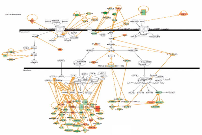
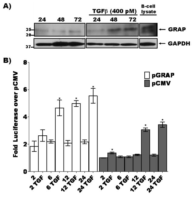
The aim of this study was to define novel mediators of tubule injury in diabetic kidney disease. For this, we used state-of-the-art proteomic methods combined with a label-free quantitative strategy to define protein expression differences in kidney tubules from transgenic OVE26 type 1 diabetic and control mice. The analysis was performed with diabetic samples that displayed a pro-fibrotic phenotype. We have identified 476 differentially expressed proteins. Bioinformatic analysis indicated several clusters of regulated proteins in relevant functional groups such as TGF-beta signaling, tight junction maintenance, oxidative stress, and glucose metabolism. Mass spectrometry detected expression changes of four physiologically relevant proteins were confirmed by immunoblot analysis. Of these, the Grb2-related adaptor protein (GRAP) was up-regulated in kidney tubules from diabetic mice and fibrotic kidneys from diabetic patients, and subsequently confirmed as a novel component of TGF-beta signaling in cultured human renal tubule cells. Thus, indicating a potential novel role for GRAP in TGF-beta-induced tubule injury in diabetic kidney disease. Although we targeted a specific disease, this approach offers a robust, high-sensitivity methodology that can be applied to the discovery of novel mediators for any experimental or disease condition.


Figures

Figure 1
Figure 1. Enhanced Pro-fibrotic signaling phenotype in renal tubules of OVE26 diabetic mice
Panel of immunoblot analyses showing increased TGF-β, GSK3β, phospho-GSK3β, and C/EBPα and decreased β-catenin levels in renal tubule extracts from OVE26 diabetic mice as compared with samples from background control mice. This expression profile is indicative of a pro-fibrotic signaling phenotype in the diabetic tubule samples. Importantly, these cellular extracts were used for quantitative 2D-LC-MS/MS analysis.
Figure 2
Figure 2. Validation of selected LC-MS/MS results
A) Quantitative LC-MS/MS differences for two key metabolic enzymes transketolase (TKT) and isocitrate dehydrogenase 3 (NAD+)-γ (IDH3) and the oxidative stress response protein DJ-1. B) Immunoblot validation. C) Densitometry shows agreement with LC-MS/MS abundance differences (* indicates p<0.05 for TKT, IDH3 or DJ-1 normalized to GAPDH or Actin loading control in diabetic tubule extracts versus control tubule extracts).
Figure 3
Figure 3. Ingenuity Pathways Knowledge Base bioinformatic analysis shows integration of candidate proteins into canonical TGF-β and BMP signaling pathways
Highlighted in green indicates down-regulated and orange indicates up-regulated in OVE26 diabetic mouse tubules.
Figure 4
Figure 4. Grb2-related adaptor protein (GRAP) expression is elevated in renal tubules from type 1 transgenic diabetic mice and diabetic patients with renal fibrosis
A) Quantitative 2D-LC-MS/MS analysis indicates that GRAP is enhanced in renal tubules from OVE26 type 1 diabetic mice, as compared with samples from control mice B) Immunoblot analysis confirms that the 25 kDa band corresponding to GRAP is increased in diabetic tubule extracts as compared to control, lower panel B shows densitometry where * indicates p<0.05 for GRAP normalized to GAPDH loading control in diabetic tubule extracts versus control tubule extracts. C) Representative immunohistochemistry (IHC) shows increased cortical tubule expression of GRAP in kidney sections from human diabetic patients that have tubulointerstitial fibrosis, as indicated by trichrome staining. In total, biopsy sections were collected from 2 diabetic and 3 normal patients. Both diabetic sections exhibited increased GRAP tubule immunostaining correlated with increased trichrome staining while 2 normal sections showed low basal GRAP levels and normal trichrome staining, one normal section exhibited increased GRAP immunostaining and increased trichrome staining.
Figure 5
Figure 5. GRAP is induced by TGF-β and potentiates the TGF-β induced SMAD response in cultured human proximal tubule HK11 cells
A) TGF-β induces expression of GRAP in cultured human proximal tubule cells. HK11 cells were treated with 400 pM TGF-β for 24, 48 and 72 hours cells harvested and lysates were fractionated by 4–15% gradient SDS-PAGE and immunoblotted for GRAP. GRAP levels increased at 24 hr post TGF-β treatment compared to unstimulated condition. B) SMAD-Luciferase reporter and either pCMV control vector (gray bars) or pCMV-GRAP (white bars) were transfected into HK11 cells and incubated with or without 400 pM TGF-β over a 2–24 hour time course then lysed and luciferase activity was measured as a function of TGF-β activity (p<0.05 indicated by * for paired comparison to 2 hour untreated controls either pGRAP or pCMV transfected).
Figure 6
Figure 6. GRAP promotes the expression of the extracellular matrix protein fibronectin in cultured human proximal tubule HK11 cells
A) HK11 cells were transfected with pGRAP (lanes 3 and 4) or pCMV control vector (lane 5) or mock transfected (lane 1 and 2). Following overnight transfection, cells representing lanes 2 and 4 were incubated with recombinant TGF-β for 20 hours. The other cells were untreated. Cell lysates were prepared, separated by 4–15% gradient SDS-PAGE, and immunoblotted for fibronectin and GAPDH as a loading control. pGRAP potentiated TGFβ induced accumulation of fibronectin. B) HK11 cells transfected with pGRAP-GFP show that GRAP localizes to the nucleus and induces secretion of fibronectin and potentiates the effects of TGFβ.
Figure 7
Figure 7. Model of GRAP regulation of TGF-β-induced tubulointerstitial fibrosis in diabetic nephropathy (DN)
Sustained diabetes can lead to renal tubule injury and concurrent TGF-β upregulation [47]. This results in exacerbation (striped arrows) of TGF-β-induced accumulation of extracellular matrix in the tubule interstitial space. Our data supports a positive feedback model were TGF-β activates increased expression of Grb2-related adaptor protein (GRAP) which enhances TGF-β SMAD driven transcription (black arrow) leading to elevated levels of fibronectin in renal proximal tubule cells.

References

    1. Molitch ME, et al. Nephropathy in diabetes. Diabetes Care. 2004;27(Suppl 1):S79–83. - PubMed
    1. Xue JL, et al. Forecast of the number of patients with end-stage renal disease in the United States to the year 2010. J Am Soc Nephrol. 2001;12(12):2753–8. - PubMed
    1. Lewis EJ, et al. Renoprotective effect of the angiotensin-receptor antagonist irbesartan in patients with nephropathy due to type 2 diabetes. N Engl J Med. 2001;345(12):851–60. - PubMed
    1. Nath KA. The tubulointerstitium in progressive renal disease. Kidney Int. 1998;54(3):992–4. - PubMed
    1. Cravatt BF, Simon GM, Yates JR., 3rd The biological impact of mass-spectrometry-based proteomics. Nature. 2007;450(7172):991–1000. - PubMed

Publication types

Substances