. 2010 Feb;134(2):121-9.
doi: 10.1016/j.clim.2009.09.005.

Monitoring of antigen-specific CD8 T cells in patients with type 1 diabetes treated with antiCD3 monoclonal antibodies

Affiliations

Monitoring of antigen-specific CD8 T cells in patients with type 1 diabetes treated with antiCD3 monoclonal antibodies

Simona Cernea et al. Clin Immunol. 2010 Feb.

Abstract

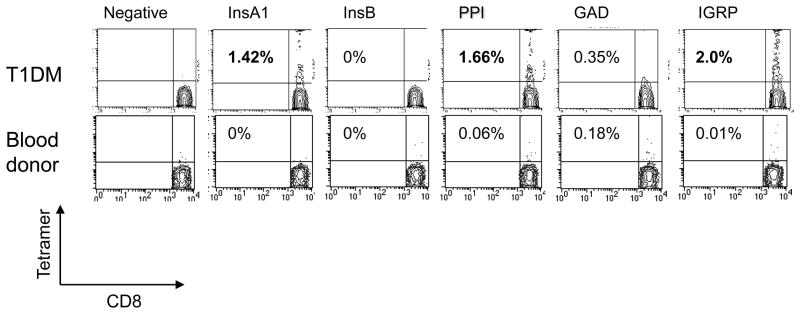
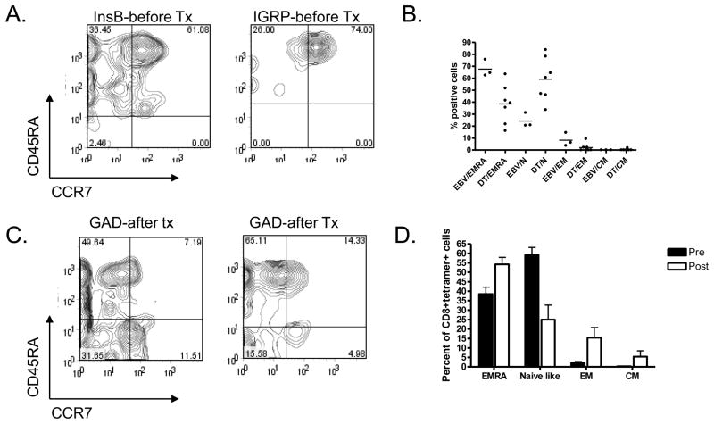
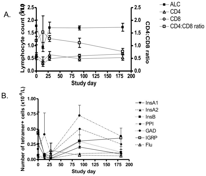
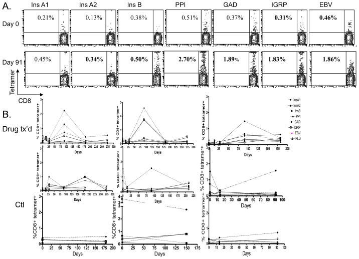
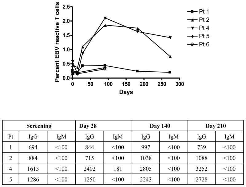
The way in which anti-CD3 monoclonal antibodies (mAbs) modify human immune responses in type 1 diabetes (T1DM) is not known. We prepared a panel of Class I HLA-A2.1 tetramers with peptides from diabetes-associated antigens and studied the frequency and phenotype of the cells in patients with T1DM and blood donors and in patients treated with anti-CD3 mAb (Teplizumab). More patients with T1DM showed positive staining for at least 1 tetramer using frozen and fresh samples (p<0.05). Three months following treatment with anti-CD3 mAb, the proportion of GAD65- and InsB-peptide reactive CD8+ T cells increased (p<0.05). The phenotype of these cells was modulated from naïve to effector memoryRA+. We concludethat Class I MHC tetramers can identify antigen specific CD8+ T cells in patients with T1DM. The frequency of certain specificities increases after treatment with anti-CD3 mAb. Their modulated phenotype may have functional consequences for their pathogenicity.


Figures

Figure 1
Figure 1. Identification of antigen specific CD8+ T cells with Class I MHC tetramers
The representative staining of fresh cells from a representative patient with T1DM and a blood donor (both HLA-A2+) are shown. The X axis shows staining with anti-CD8 mAb and the Y axis represents staining with tetramer. Electronic gates were placed around lymphocytes on the basis of forward and side scatter and around CD8+ cells. The numbers in each contour plot represents the percentage of CD8+ T cells that are tetramer+ minus the background staining with the negative tetramer (% negative tetramer: 0.06% [T1DM] and 0.03% [Blood donor], respectively). Bolded numbers represent positive staining (above threshold for positivity from staining of HLA-A2− control subjects (see Supplemental Table 1)).
Figure 2
Figure 2. Phenotype of tetramer+ cells
A: PBMC were stained with mAbs to CD8, CCR7, CD45RA, and the indicated tetramers. Electronic gates were placed around the CD8+tetramer+ cells and the expression of CCR7 (X axis) and CD45RA (Y axis) was analyzed. B: The functional phenotypes, on the basis of expression of CCR7 and CD45RA are shown for diabetes antigen specific CD8+ T cells (DT) and EBV specific T cells (EBV) from patients with T1DM. There was a significant difference in the distribution of phenotypes among the DT+ vs EBV+ T cells (p<0.001). EMRA: terminally differentiated effector memory RA+; N: naïve-like; EM: effector memory; CM: central memory; C: The expression of CCR7 and CD45RA among diabetes antigen specific T cells after treatment with anti-CD3 mAb is shown. D: The frequency of phenotypes of CD8+diabetes tetramer+ T cells before and after treatment with anti-CD3 mAb are shown. There was a significant change in the distribution of phenotypes after treatment (p<0.001).
Figure 3
Figure 3. Changes in the numbers of lymphocytes during and following treatment with Teplizumab
A: The absolute lymphocyte (ALC) (n=6), CD4, CD8 (L axis), cell counts, and CD4:CD8 ratios (R axis)(n=6) were calculated for subjects receiving Teplizumab for 14 days. B. The absolute numbers of tetramer+ cells are shown.
Figure 4
Figure 4. Changes in diabetes antigen specific CD8+ T cells with treatment with Teplizumab
A Representative staining for CD8 and tetramers from a single participant is shown before (day 0) and 3 months after (day 90) treatment with Teplizumab. The numbers refer to the percentage tetramer+ of the CD8+ T cells. Bolded numbers are considered positive staining based on threshold values shown in Supplemental Figure 1. B: Changes in the frequency of diabetes antigen specific T cells in individual patients treated with Teplizumab and untreated patients with T1DM. (tetramers: InsA1 ▼;, InsA2 ▲, Ins B ■ PPI ◆ GAD ● IGRP □, Flu ○).
Figure 5
Figure 5. Changes in EBV reactive T cells and anti-EBV immunoglobulins after treatment with Teplizumab
The changes in EBV reactive CD8+ T cells were monitored in parallel with the diabetes-antigen specific T cells. The EBV IgG and IgM titers were measured at the indicated study days (the mAb was given from days 0–14).

References

    1. Atkinson MA. ADA Outstanding Scientific Achievement Lecture 2004. Thirty years of investigating the autoimmune basis for type 1 diabetes: why can’t we prevent or reverse this disease? Diabetes. 2005;54:1253–63. - PubMed
    1. Miller BJ, Appel MC, O’Neil JJ, Wicker LS. Both the Lyt-2+ and L3T4+ T cell subsets are required for the transfer of diabetes in nonobese diabetic mice. J Immunol. 1988;140:52–8. - PubMed
    1. Christianson SW, Shultz LD, Leiter EH. Adoptive transfer of diabetes into immunodeficient NOD-scid/scid mice. Relative contributions of CD4+ and CD8+ T-cells from diabetic versus prediabetic NOD. NON-Thy-1a donors. Diabetes. 1993;42:44–55. - PubMed
    1. Katz JD, Wang B, Haskins K, Benoist C, Mathis D. Following a diabetogenic T cell from genesis through pathogenesis. Cell. 1993;74:1089–100. - PubMed
    1. Amrani A, Verdaguer J, Serra P, Tafuro S, Tan R, Santamaria P. Progression of autoimmune diabetes driven by avidity maturation of a T-cell population. Nature. 2000;406:739–42. - PubMed

Publication types

MeSH terms