. 2009 Oct 16;139(2):337-51.
doi: 10.1016/j.cell.2009.07.052.

GOLPH3 bridges phosphatidylinositol-4- phosphate and actomyosin to stretch and shape the Golgi to promote budding

Affiliations

GOLPH3 bridges phosphatidylinositol-4- phosphate and actomyosin to stretch and shape the Golgi to promote budding

Holly C Dippold et al. Cell. .

Abstract

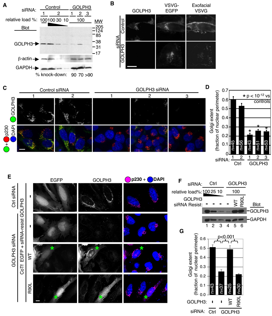
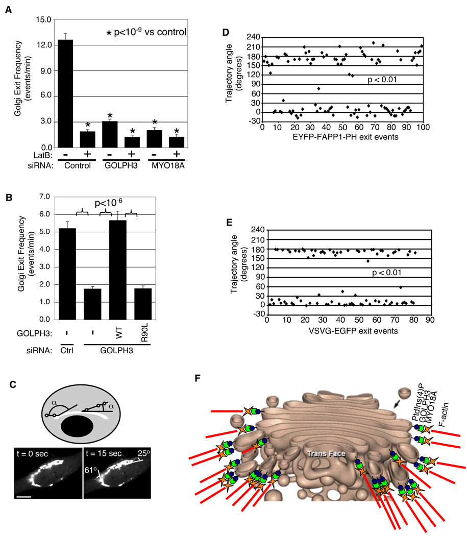
Golgi membranes, from yeast to humans, are uniquely enriched in phosphatidylinositol-4-phosphate (PtdIns(4)P), although the role of this lipid remains poorly understood. Using a proteomic lipid-binding screen, we identify the Golgi protein GOLPH3 (also called GPP34, GMx33, MIDAS, or yeast Vps74p) as a PtdIns(4)P-binding protein that depends on PtdIns(4)P for its Golgi localization. We further show that GOLPH3 binds the unconventional myosin MYO18A, thus connecting the Golgi to F-actin. We demonstrate that this linkage is necessary for normal Golgi trafficking and morphology. The evidence suggests that GOLPH3 binds to PtdIns(4)P-rich trans-Golgi membranes and MYO18A conveying a tensile force required for efficient tubule and vesicle formation. Consequently, this tensile force stretches the Golgi into the extended ribbon observed by fluorescence microscopy and the familiar flattened form observed by electron microscopy.


Figures

Figure 1
Figure 1. Proteomic screening identifies GOLPH3 as a PtdIns(4)P binding protein that requires PtdIns(4)P for Golgi localization
(A) Screening of Drosophila Gene Collection for lipid binding. Example hits correspond to previously well-validated lipid binding proteins with known binding domains: AKT (PtdIns(3,4)P2 and PtdIns(3,4,5)P3, clone SD10374); CERT (PtdIns(4)P, clone GH07688); TAPP1 (PtdIns(3,4)P2, clone SD10969); SNX29 (PtdIns(3)P and PA, clone LD35592); SARA (PtdIns(3)P, clone LD33044); Tubby (PtdIns(4,5)P2, clone GH04653); ATG18 (PtdIns(3,5)P2, PtdIns(3)P, clone LD32381). Screen also identified unknown lipid binding proteins. Shown is PtdIns(4)P binding of GOLPH3 (clone LD23816). Yeast (Vps74p) and human orthologs also bind PtdIns(4)P. (B) Depletion of PtdIns(4)P results in dissociation of GOLPH3 from the Golgi. HeLa cells expressing EGFP-tagged phosphatase Sac1-K2A (constitutively at Golgi, Rohde et al., 2003) or EGFP (control) stained for endogenous GOLPH3 (red), TGN marker p230 (yellow), and DAPI (blue). In controls and untransfected cells (*), GOLPH3 and p230 co-localize at the Golgi, but EGFP-Sac1-K2A renders GOLPH3 staining diffuse and distinct from p230. (C) Overexpression of EYFP-FAPP1-PH displaces endogenous GOLPH3 from the Golgi. White arrowheads indicate cells expressing EYPF-FAPP1-PH (green) in which anti-GOLPH3 staining (red) is diffuse vs adjacent untransfected cells. (D) S. cerevisiae expressing temperature sensitive allele pik1-83ts, lose PtdIns(4)P upon shift to 37°C. In wild type, GFP-Vps74p remains colocalized with late Golgi marker Sec7p-dsRed at both temperatures. In pik1-83ts, GFP-Vps74p dissociates from the Golgi upon shift to 37°C (also see Movie S2). (Figure 1–Figure 5 and Figure 7, scale bars=10 µm)
Figure 2
Figure 2. Identification of PtdIns(4)P binding domain and pocket required for Golgi localization of GOLPH3
(A, B, C) Truncations of Drosophila GOLPH3 show PtdIns(4)P binding in vitro co-maps with Golgi localization. (A) Segments indicated expressed in vitro for lipid blot or (C) as EGFP-fusions in HEK 293 cells for fluorescence microscopy. (B) Expression confirmed by Western blot for EGFP. (D) Lipid vesicle binding shows specificity of wild type GOLPH3 for PtdIns(4)P. R90L and R171A/R174L mutants do not bind. p40phox-PX control binds PtdIns(3)P. (V=vesicle, S=soluble) (E) In HeLa, R90L and R171A/R174L mutations of GOLPH3 (magenta) mislocalize to cytosol, but coexpressed wild type EGFP-GOLPH3 (green) remains at the Golgi as marked by TGN46 (red). DAPI in blue. (F) Western blot showing coexpression of 3xHA-tagged GOLPH3 mutants and EGFP-GOLPH3.
Figure 3
Figure 3. GOLPH3 is required for trafficking and extended ribbon morphology of the Golgi
(A) Western blot of HeLa lysates shows GOLPH3 knocked down 70–90% by each specific siRNA. GOLPH3 antiserum recognizes a single band at 34 kDa in control lysates. Decreasing amounts of control lysate loaded to allow quantification. Blots for β-actin and glyceraldehyde-3-phosphate dehydrogenase (GAPDH) verify equal loading. (B) Trafficking of ts045-VSVG-EGFP, through Golgi to PM impaired by knockdown of GOLPH3. HeLa cells serially transfected with GOLPH3 or control siRNA, then ts045-VSVG-EGFP expression vector, incubated at 40°C overnight, then shifted to 32°C for 2 hours. Arrival at PM determined with antibody to extracellular domain applied to unpermeabilized cells (exofacial VSVG). (C) Knockdown of GOLPH3 causes condensation of Golgi ribbon. Control siRNA transfected cells show normal extended Golgi ribbon detected by GOLPH3 (green) and p230 (red). DAPI in blue. GOLPH3 siRNA reduces GOLPH3 and causes condensation of Golgi (p230). Imaging parameters identical in all. (D) Quantification of Golgi extent (Golgi length relative to nuclear perimeter, mean/SEM graphed) demonstrates significant change (p<10−12, t-test). (E) Expression of siRNA-resistant GOLPH3 (expressed near endogenous level, cotransfected with EGFP) rescues Golgi ribbon, validating GOLPH3 siRNA specificity. Expression of siRNA-resistant GOLPH3 PtdIns(4)P binding pocket mutant does not rescue Golgi ribbon phenotype. Asterisks indicate transfected cells. (F) Western blot shows knockdown of GOLPH3 (lane 4) and restoration by siRNA-resistant wild type (lane 5) and R90L (lane6) GOLPH3. Blot for GAPDH shows equal loading. (G) Quantification of Golgi extent (mean/SEM) demonstrates significant rescue by wild type, but not R90L GOLPH3 (p<0.001 for indicated comparisons, t-test).
Figure 4
Figure 4. GOLPH3 links the Golgi to F-actin via MYO18A
(A) HeLa cells treated with control siRNA and DMSO show normal extended Golgi morphology (p230, green and GOLPH3, yellow) and normal F-actin (Texas red phalloidin, magenta). Control siRNA + LatB causes loss of F-actin (stress fibers and peripheral actin) and Golgi condensation, but GOLPH3 remains at Golgi. GOLPH3 siRNA + DMSO causes loss of GOLPH3, Golgi condensation, but F-actin remains normal. Combined GOLPH3 siRNA + LatB causes loss of F-actin and GOLPH3, with Golgi condensation. (B) Measurement of Golgi extent (from A) indicates no additional effects on Golgi seen in combining GOLPH3 siRNA and LatB. (C) Western blot shows MYO18A coIPs specifically with GOLPH3 even when actin depolymerized by LatB (Figure S13). Golgi-localized myosins, MYO2B and MYO6, and MRCKβ did not coIP with GOLPH3. (D) Proposed model: GOLPH3 binds PtdIns(4)P and MYO18A, linking Golgi to cytoskeleton. (E) Purified, bacterial expressed GOLPH3 (but not GST) binds His-SUMO-tagged N-terminal and Middle fragments of MYO18A but not C-terminal fragment or His-SUMO alone bound to Ni beads. (F) MYO18A (green) colocalizes with GOLPH3 (red) and p230 (magenta) in HeLa cells. DAPI in blue. Knockdown of MYO18A shows specificity of MYO18A antibody for IF. Knockdown of GOLPH3 causes loss of MYO18A from Golgi.
Figure 5
Figure 5. MYO18A knockdown phenocopies GOLPH3 compact Golgi phenotype
(A) Western blot of HeLa lysates shows MYO18A knocked down >90% by each specific siRNA. GAPDH blot verifies equal loading. (B) Knockdown of MYO18A with each specific siRNA results in condensed Golgi shown by IF to GOLPH3 (green) and p230 (red). DAPI in blue. Similar results seen in HeLa (shown) and HEK 293 cells. (C) Quantification of Golgi extent (length relative to nuclear perimeter, mean/SEM graphed) demonstrates significant compaction of Golgi (p<10−10, t-test). (D) GFP-tagged wild type mouse MYO18A, but not ATP-binding mutant nor EGFP alone, rescues Golgi phenotype of MYO18A knockdown in HeLa cells, shown by IF to GOLPH3 and p230 (magenta). (E) Quantification of Golgi extent (mean/SEM graphed) demonstrates significant rescue of Golgi morphology by wild type MYO18A, but not ATP-binding mutant (p<0.02 for indicated comparisons, t-test).
Figure 6
Figure 6. Knockdown of GOLPH3 or MYO18A produces dilated Golgi cisternae
(A) Electron micrographs of negative control, GOLPH3, and MYO18A siRNA-treated HeLa cells (two different oligos for each). Control cells show flat Golgi cisternae arranged in neat stacks. Knockdown of GOLPH3 or MYO18A (verified by Western blot of parallel samples) results in dilated, frequently completely aberrant, Golgi cisternae, especially on trans face (see text). (scale bar=200 nm) (B) Cisternae in each stack divided into the cis-medial and medial-trans halves and thickness of each cisterna measured. Differences between control and knockdown highly significant for medial-trans (p<10−4, t-test, mean/SEM graphed).
Figure 7
Figure 7. GOLPH3/MYO18A/F-actin are necessary for Golgi vesiculation
(A) Knockdown of GOLPH3, MYO18A, or depolymerization of actin impair Golgi vesicle exit. HeLa cells expressing low levels of EYFP-FAPP1-PH to mark PtdIns(4)P-rich membranes imaged live before and after LatB treatment. Vesicles or tubules exiting Golgi counted (10–15 cells per siRNA, counting 100–1500 exit events per siRNA, pooling two experiments). Differences from control highly significant (p<10−9, t-test). (B) siRNA-resistant wild-type GOLPH3 rescues Golgi trafficking defect, but not the R90L mutant defective in binding PtdIns(4)P. Differences with p<10−6 by t-test (20–30 cells each, counting 900–2400 exit events, pooling two experiments). (C) Measurement of exit angle of tubules/vesicles from Golgi. Live imaging of EYFP-FAPP1-PH shows two PtdIns(4)P-rich tubules leaving Golgi (see also Movie S4) and measurement of initial trajectory angles relative to direction of Golgi ribbon. (D) Initial trajectories of PtdIns(4)P-rich tubules or vesicles plotted for 99 exit events collected from independent imaging of 8 HeLa cells. Clustering around 0° and 180° highly significant (p<0.01, Kolmogorov-Smirnov test). (D) Initial trajectories of vesicles and tubules carrying ts045-VSVG-GFP cluster near 0° and 180°, parallel to Golgi ribbon (p<0.01, Kolmogorov-Smirnov test). (E) Diagram of Golgi (used with permission (Davidson, 2004)) superimposed with Model: GOLPH3 binds PtdIns(4)P and links to MYO18A, transducing a tensile force necessary for Golgi vesiculation, contributing to its flattened form (see also Figure 4D).

References

    1. Allan VJ, Thompson HM, McNiven MA. Motoring around the Golgi. Nat. Cell Biol. 2002;4:E236–E242. - PubMed
    1. Audhya A, Foti M, Emr SD. Distinct roles for the yeast phosphatidylinositol 4-kinases, Stt4p and Pik1p, in secretion, cell growth, and organelle membrane dynamics. Mol. Biol. Cell. 2000;11:2673–2689. - PMC - PubMed
    1. Barr FA, Warren G. Disassembly and reassembly of the Golgi apparatus. Semin. Cell Dev. Biol. 1996;7:505–510.
    1. Beh CT, Cool L, Phillips J, Rine J. Overlapping functions of the yeast oxysterol-binding protein homologs. Genetics. 2001;157:1117–1140. - PMC - PubMed
    1. Bell AW, Ward MA, Blackstock WP, Freeman HNM, Choudhary JS, Lewis AP, Chotai D, Fazel A, Gushue JN, Paiement J, Palcy S, Chevet E, Lafrenière-Roula M, Solari R, Thomas DY, Rowley A, Bergeron JJM. Proteomics characterization of abundant Golgi membrane proteins. J. Biol. Chem. 2001;276:5152–5165. - PubMed

Publication types