. 2009 Oct 22;6(4):343-53.
doi: 10.1016/j.chom.2009.09.002.

Divergence of macrophage phagocytic and antimicrobial programs in leprosy

Affiliations

Divergence of macrophage phagocytic and antimicrobial programs in leprosy

Dennis Montoya et al. Cell Host Microbe. .

Abstract

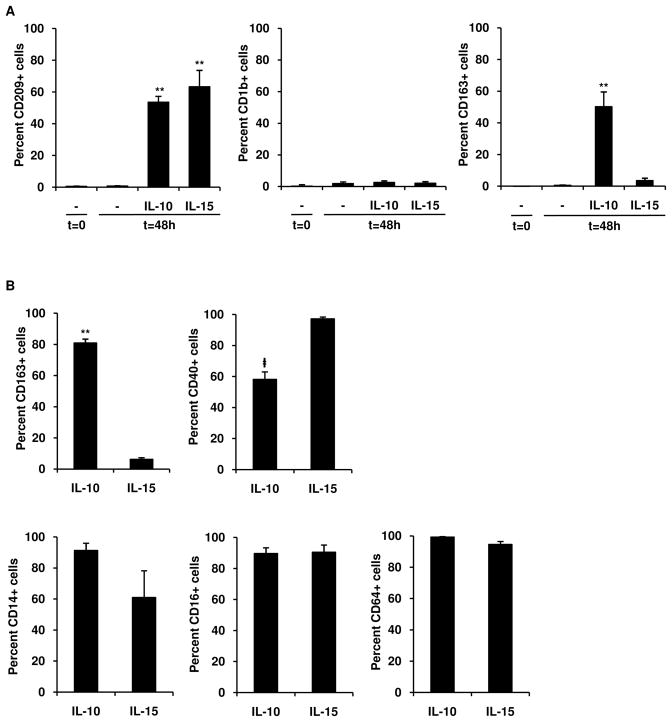
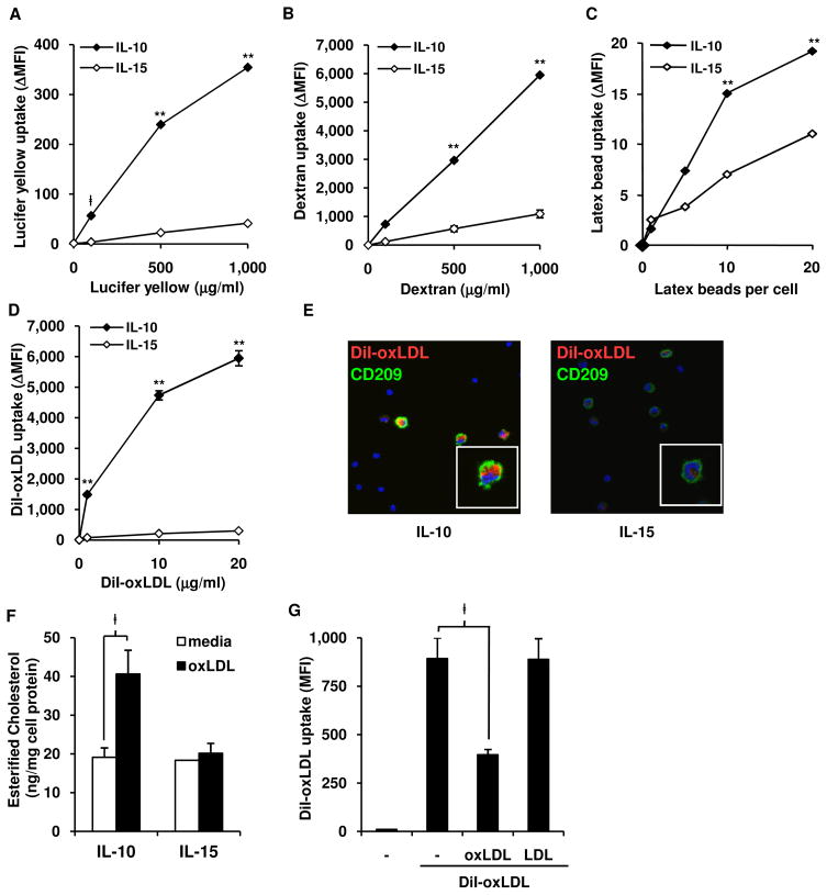
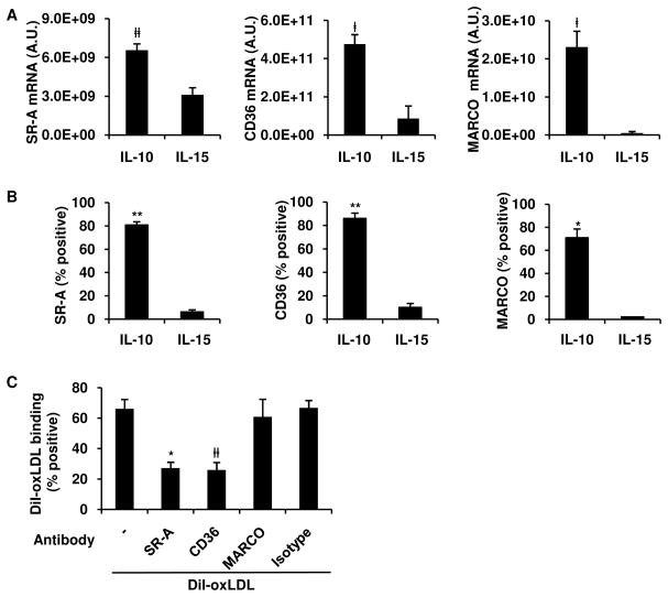
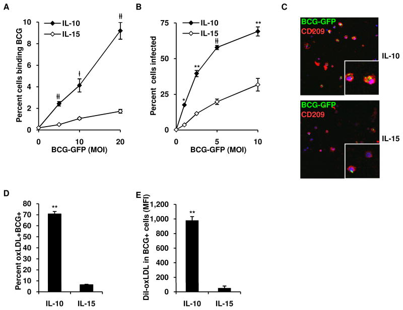
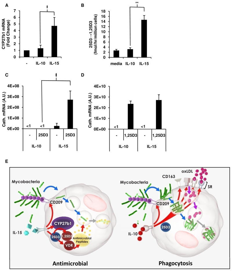
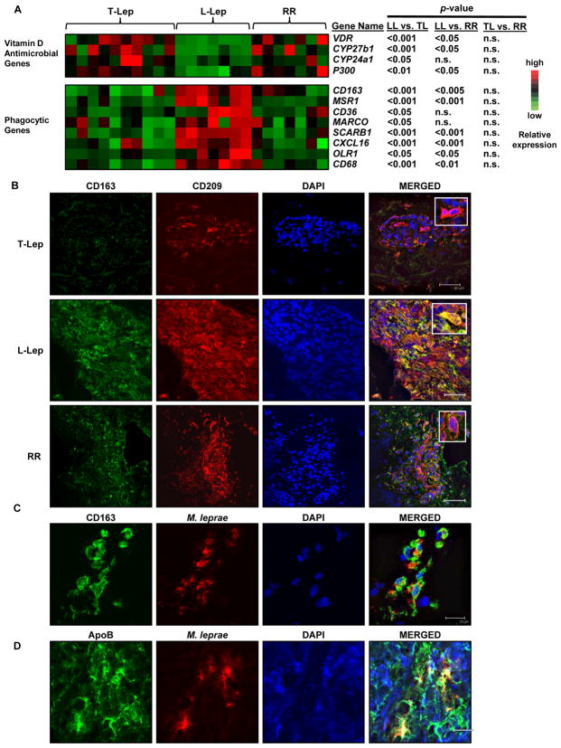
Effective innate immunity against many microbial pathogens requires macrophage programs that upregulate phagocytosis and direct antimicrobial pathways, two functions generally assumed to be coordinately regulated. We investigated the regulation of these key functions in human blood-derived macrophages. Interleukin-10 (IL-10) induced the phagocytic pathway, including the C-type lectin CD209 and scavenger receptors, resulting in phagocytosis of mycobacteria and oxidized low-density lipoprotein. IL-15 induced the vitamin D-dependent antimicrobial pathway and CD209, yet the cells were less phagocytic. The differential regulation of macrophage functional programs was confirmed by analysis of leprosy lesions: the macrophage phagocytosis pathway was prominent in the clinically progressive, multibacillary form of the disease, whereas the vitamin D-dependent antimicrobial pathway predominated in the self-limited form and in patients undergoing reversal reactions from the multibacillary to the self-limited form. These data indicate that macrophage programs for phagocytosis and antimicrobial responses are distinct and differentially regulated in innate immunity to bacterial infections.


Figures

Figure 1
Figure 1. IL-10 differentiates monocytes into CD209+CD163+ MΦ
(A) Human peripheral monocytes were harvested at 0 h or stimulated for 48 h with IL-10, IL-15, or media and labeled with specific antibodies. Results shown as mean ± SEM (n ≥ 4). **p < 0.001 versus media (t = 48 h). (B) IL-10 and IL-15 derived MΦ were double-labeled with antibodies against CD209 and specific markers. Results represent mean ± SEM (n ≥ 4) percent of CD209+ cells positive for indicated antibody. p < 0.05 **p < 0.001 versus IL-15 derived MΦ.
Figure 2
Figure 2. IL-10 derived MΦ have enhanced endocytic activity and develop into foam cell MΦ
(A–D) Endocytosis assays comparing IL-10 and IL-15 programmed MΦ with (A) lucifer yellow (B) FITC-labeled dextran, (C) fluorescent latex beads, or (D) DiI-oxLDL, at indicated concentrations. Cells assayed for uptake at 37°C or binding at 4°C, labeled for CD209, data represented as net intracellular mean florescence intensity (ΔMFI=MFIuptake-MFIbinding) of indicated dye in CD209+ cells. Data represent the mean (n ≥ 3) ± SEM p < 0.05, **p < 0.001 versus IL-15 derived MΦ. (E) Confocal images of cells cultured and labeled as in (D). DiI-oxLDL, red; CD209, green; DAPI, blue. (F) IL-10 or IL-15 derived MΦ were incubated with unlabeled oxLDL, lipids extracted and mean ± SEM (n = 3) of esterified cholesterol shown, normalized to amount of cell protein. (G) Uptake of DiI-oxLDL was competed against excess amounts (30X of DiI-oxLDL) of unlabeled oxLDL, LDL or media and analyzed as in (D). Data represent the mean (n = 3) ± SEM p < 0.05, *p < 0.005, **p < 0.001 versus IL-15 derived MΦ.
Figure 3
Figure 3. SR-A and CD36 mediate oxLDL uptake in IL-10 programmed MΦ
(A–B) Monocytes were stimulated with IL-10 or IL-15 and scavenger receptor expression assessed by qPCR (A) after 24 r or surface protein expression (B) after 48 h on CD209+ cells by flow cytometry. Results show as mean ± SEM (n ≥ 3). (C) Blocking antibodies against SR-A, CD36, MARCO, or isotype were pre-incubated with of IL-10 programmed MΦ and binding of DiI-oxLDL assayed. All results shown as mean ± SEM (n ≥ 3). p < 0.05, #p < 0.01 *p < 0.005, **p < 0.001 versus IL-15 treated monocytes.
Figure 4
Figure 4. IL-10 derived MΦ coordinately phagocytose mycobacteria and oxLDL
(A) Binding of BCG-GFP by IL-10 and IL-15 programmed MF (B) Phagocytosis of BCG-GFP by IL-10 and IL-15 programmed MF. Results shown as mean ± SEM (n = 4) percent of CD209+ cells positive for BCG. (C) Confocal images of MΦ cultured as in (B) (BCG-GFP, green; CD209, red; DAPI, blue). (D–E) Coordinate uptake of oxLDL and BCG-GFP by IL-10 or IL-15 programmed MΦ, (D) percent of CD209+ cells positive for oxLDL and BCG. (E) Amount of DiI-oxLDL in CD209+ cells infected with BCG. Results show as mean ± SEM (n = 3). p < 0.05, #p < 0.01, *p < 0.005, **p < 0.001 versus controls.
Figure 5
Figure 5. IL-15 vs. IL-10 differentially programs the vitamin D antimicrobial pathway in MΦ
(A) CYP27b1 expression measured by qPCR in monocytes stimulated with IL-10, IL-15 or media. Results shown as mean ± SEM (n = 3) fold change normalized to media stimulated cells. (B) IL-10 or IL-15 derived MΦ cultured with radiolabeled 25D3 and rate of conversion to 1,25D3 measured by HPLC. (C–D) Expression of cathelicidin mRNA after culturing IL-10 and IL-15 programmed MΦ with 25D3 (C) or 1,25D3 (D) vitamin D. All data represented as mean ± SEM (n = 3). p < 0.05, **p < 0.001. (E) Proposed model of divergence of phagocytosis and antimicrobial programs in MΦ. IL-10 induces a scavenger receptor program resulting in enhanced phagocytosis resulting in microbial persistence, while IL-15 induces the vitamin D mediated antimicrobial program resulting in microbial killing.
Figure 6
Figure 6. Dynamics of the clinical response in leprosy patients reflects the phenotype and function of the IL-10 and IL-15 derived MΦ programs
(A) Gene expression analysis of leprosy skin biopsies for vitamin D antimicrobial pathway and phagocytic program. Each column represents one donor. n.s. not significant. (B) Phenotype of MΦ in human leprosy skin lesions: L-lep and T-lep skin lesions were labeled for CD163 (green), CD209 (red), and DAPI (blue). Scale bar 40μm. (C–D) CD163+ MΦ in L-lep lesions labeled for accumulation of oxLDL and mycobacteria, by antibodies against (C) M. leprae or (D) ApoB. Representative data of at least three patients used for each groups. Scale bar 20μm.

References

    1. Arredouani MS, Palecanda A, Koziel H, Huang YC, Imrich A, Sulahian TH, Ning YY, Yang Z, Pikkarainen T, Sankala M, Vargas SO, Takeya M, Tryggvason K, Kobzik L. MARCO is the major binding receptor for unopsonized particles and bacteria on human alveolar macrophages. J Immunol. 2005;175:6058–6064. - PubMed
    1. Babu S, Kumaraswami V, Nutman TB. Alternatively activated and immunoregulatory monocytes in human filarial infections. J Infect Dis. 2009;199:1827–1837. - PMC - PubMed
    1. Backman KA, Guyre PM. Gamma-interferon inhibits Fc receptor II-mediated phagocytosis of tumor cells by human macrophages. Cancer Res. 1994;54:2456–2461. - PubMed
    1. Barnetson RS, Bjune G, Pearson JMH, Kronvall G. Cell mediated and humoral immunity in “reversal reactions”. Int J Lepr. 1976;44:267–273. - PubMed
    1. Bjune G, Barnetson RS, Ridley DS, Kronvall G. Lymphocyte transformation test in leprosy: correlation of the response with inflammation of lesions. Clin Exp Immunol. 1976;25:85–94. - PMC - PubMed

Publication types

Associated data

LinkOut - more resources