. 2009 Dec 4;284(49):33876-82.
doi: 10.1074/jbc.M109.050989. Epub 2009 Oct 16.

Substrate-induced assembly of Methanococcoides burtonii D-ribulose-1,5-bisphosphate carboxylase/oxygenase dimers into decamers

Affiliations

Substrate-induced assembly of Methanococcoides burtonii D-ribulose-1,5-bisphosphate carboxylase/oxygenase dimers into decamers

Hernán Alonso et al. J Biol Chem. .

Abstract

Like many enzymes, the biogenesis of the multi-subunit CO(2)-fixing enzyme ribulose-1,5-bisphosphate (RuBP) carboxylase/oxygenase (Rubisco) in different organisms requires molecular chaperones. When expressed in Escherichia coli, the large (L) subunits of the Rubisco from the archaeabacterium Methanococcoides burtonii assemble into functional dimers (L(2)). However, further assembly into pentamers of L(2) (L(10)) occurs when expressed in tobacco chloroplasts or E. coli producing RuBP. In vitro analyses indicate that the sequential assembly of L(2) into L(10) (via detectable L(4) and L(6) intermediates) occurs without chaperone involvement and is stimulated by protein rearrangements associated with either the binding of substrate RuBP, the tight binding transition state analog carboxyarabinitol-1,5-bisphosphate, or inhibitory divalent metal ions within the active site. The catalytic properties of L(2) and L(10) M. burtonii Rubisco (MbR) were indistinguishable. At 25 degrees C they both shared a low specificity for CO(2) over O(2) (1.1 mol x mol(-1)) and RuBP carboxylation rates that were distinctively enhanced at low pH (approximately 4 s(-1) at pH 6, relative to 0.8 s(-1) at pH 8) with a temperature optimum of 55 degrees C. Like other archaeal Rubiscos, MbR also has a high O(2) affinity (K(m)(O(2)) = approximately 2.5 microM). The catalytic and structural similarities of MbR to other archaeal Rubiscos contrast with its closer sequence homology to bacterial L(2) Rubisco, complicating its classification within the Rubisco superfamily.


Figures

FIGURE 1.
FIGURE 1.
Ligand induced sequential assembly of MbR L2 units at room temperature into stable decamers. Nondenaturing PAGE (A, C, and E) and nanoESI-MS (B, D, and F) of Rubisco complexes with the corresponding masses derived from the m/z of the ions shown in brackets. A, separation of purified tobacco L8S8 Rubisco (twt), tobacco produced R. rubrum L2 Rubisco from cmtrL1 (tRr) (24) and E. coli purified L2 MbR (EMbR) (supplemental Fig. 2). B, nanoESI-MS of the denatured EMbR monomer (5% (v/v) formic acid) and native dimer (0.1 m ammonium acetate, pH 7). The major ion in the spectrum of the monomer is at m/z 3524.8 corresponding to [MbR+15H]15+, and for the dimer it is 5564.9 corresponding to [MbR+19H]19+. C–F, induced assembly of CO2-Mg2+ activated MbR L2 units (final concentration, 3.8 μm) into L4, L6, and L10 complexes by 1 mm RuBP (C) and 1 mm CABP (E) over time and the corresponding nanoESI-MS of nonactivated MbR after 30 min of incubation with 100 μm RuBP (D) or CABP (F). Where RuBP or CABP are present (D and F), the peaks in nanoESI mass spectra are broad, making it difficult to determine precise binding stoichiometries. The data were, however, consistent with the binding of one RuBP or CABP/monomer. m, marker proteins (sizes shown).
FIGURE 2.
FIGURE 2.
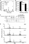
Influence of activation with Mg2+, Ca2+, or Co2+ on MbR activity, sugar-phosphate binding, and structure. A, recovery of RuBP-dependent 14CO2 activity over time relative to the activity of the Mg2+ activated MbR after 8 min. B, recovery of bound [14C]CABP or [3H]RuBP after activating decarbamylated L2 MbR for 15 min at 25 °C with 25 mm NaHCO3 and either 20 mm MgCl2 or 20 mm CaCl2 before incubating with 2 μm of [14C]CABP (1.6 MBq·μmol−1, white bars) or [3H]RuBP (16 MBq·μmol−1, black bars) for 15 min and resolving the amount bound to MbR by Superdex G50 fine gel filtration (25). The values ± S.D. are the averages of duplicate (n = 2) samples. C, nondenaturing PAGE analysis of the influence of activation with Mg2+, Ca2+, or Co2+, with or without CABP, on the assembly of L2 MbR into L10 after 20 min at 25 °C. D, nanoESI-MS of MbR in 100 mm ammonium acetate, pH 7, incubated for 30 min at 2 °C with 100 μm of MgCl2, CaCl2, or CoCl2.
FIGURE 3.
FIGURE 3.
RuBP stimulates assembly of L10 MbR in vivo. A, comparative organization of the plastomes of wild-type tobacco (twt), the tobacco master line cmtrL1 (24) (tRr), and transplastomic tobacco producing MbR (tMbR). Plasmid pLevMbiiL was biolistically transformed into cmtrL1 where the flanking plastome sequence in pLevMbiiL (indicated by dotted lines with the numbering relative to the Nicotiana tabacum plastome sequence; GenBankTM accession number Z00044) directed the homologous replacement of the R. rubrum L2 Rubisco gene (cmrbcM), psbA 3′-untranslated region (T), and loxP (triangle) sequences with a bicistronic gene encompassing rbcLMb, 216 bp of the tobacco rbcL 3′-untranslated region (T), a promoter-less aadA (marker) gene, and rps16 3′-untranslated region (t) sequence. B, SDS-PAGE and immunoblot analysis of 10 μg of leaf protein from the representative tobacco lines (noting the N terminus of MbR in tMbR, MSPQTETKASVGF, varies from the E. coli purified MbR sequence, MSLIYEDLV). C, nondenaturing PAGE of the samples from B and the purified H6Ub-MbR expressed in RuBP producing E. coli-PRK cells before (UbL10) and after (L10) removal of the H6Ub fusion (see supplemental Fig. 2). L8S8, Form I tobacco Rubisco; m, marker proteins (sizes shown).
FIGURE 4.
FIGURE 4.
The L2 to L10 transition is reversible. A, nondenaturing PAGE analysis of purified L10 MbR (final concentration, 7.5 pm) before (0 h) and after 24 h of dialysis in buffer (50 mm HEPES-NaOH, pH 7.0, 20 mm EDTA) at 4 or 22 °C. After 24 h at 22 °C, the dialyzed MbR was incubated with 20 mm MgCl2 and 1 mm CABP for 30 min at 22 °C prior to electrophoresis. B, schematic proposing a reversible assembly process of L2 MbR via possible L4 and L6 intermediates into a pentameric ring.
FIGURE 5.
FIGURE 5.
Influence of pH and temperature on MbR activity. Comparative preference of L10 MbR for low pH (A) and high temperature (B). The RuBP-carboxylase activity of L2 (○) and L10 (●) MbR at 25 and 55 °C are alike. All of the assays were performed anaerobically.

References

    1. Young J. C., Agashe V. R., Siegers K., Hartl F. U. (2004) Nat. Rev. Mol. Cell Biol. 5, 781–791 - PubMed
    1. Goloubinoff P., Christeller J. T., Gatenby A. A., Lorimer G. H. (1989) Nature 342, 884–889 - PubMed
    1. Andersson I., Backlund A. (2008) Plant Physiol. Biochem. 46, 275–291 - PubMed
    1. Brinker A., Pfeifer G., Kerner M. J., Naylor D. J., Hartl F. U., Hayer-Hartl M. (2001) Cell 107, 223–233 - PubMed
    1. Viitanen P. V., Lubben T. H., Reed J., Goloubinoff P., O'Keefe D. P., Lorimer G. H. (1990) Biochemistry 29, 5665–5671 - PubMed

Publication types

LinkOut - more resources