. 2009 Dec;191(24):7587-96.
doi: 10.1128/JB.01110-09. Epub 2009 Oct 16.

Roles of the Bacillus anthracis spore protein ExsK in exosporium maturation and germination

Affiliations

Roles of the Bacillus anthracis spore protein ExsK in exosporium maturation and germination

Kari M Severson et al. J Bacteriol. 2009 Dec.

Abstract

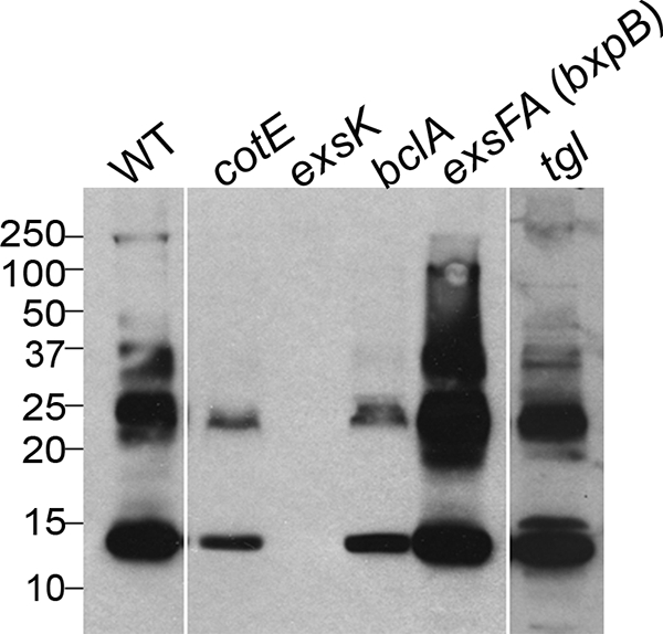
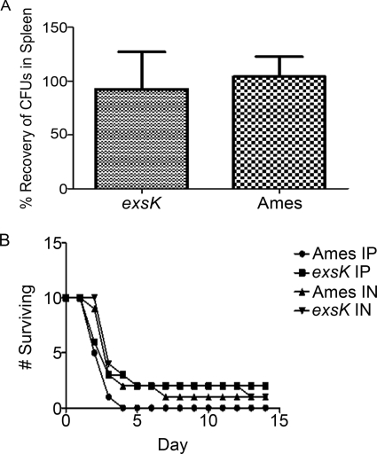
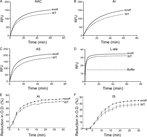
The Bacillus anthracis spore is the causative agent of the disease anthrax. The outermost structure of the B. anthracis spore, the exosporium, is a shell composed of approximately 20 proteins. The function of the exosporium remains poorly understood and is an area of active investigation. In this study, we analyzed the previously identified but uncharacterized exosporium protein ExsK. We found that, in contrast to other exosporium proteins, ExsK is present in at least two distinct locations, i.e., the spore surface as well as a more interior location underneath the exosporium. In spores that lack the exosporium basal layer protein ExsFA/BxpB, ExsK fails to encircle the spore and instead is present at only one spore pole, indicating that ExsK assembly to the spore is partially dependent on ExsFA/BxpB. In spores lacking the exosporium surface protein BclA, ExsK fails to mature into high-molecular-mass species observed in wild-type spores. These data suggest that the assembly and maturation of ExsK within the exosporium are dependent on ExsFA/BxpB and BclA. We also found that ExsK is not required for virulence in murine and guinea pig models but that it does inhibit germination. Based on these data, we propose a revised model of exosporium maturation and assembly and suggest a novel role for the exosporium in germination.


Figures

FIG. 1.
FIG. 1.
Fluorescence microscopy of B. anthracis (Sterne) sporangia. Phase-contrast (Phase) and fluorescence (GFP, Hoechst, and Merge) images of B. anthracis exsK-gfp (A, C, E, G, and I) or wild-type (WT) (B, D, F, H, and J) sporangia harvested at the indicated time points (in hours) during sporulation (t0 to t10) and visualized for GFP fluorescence and DNA staining with Hoechst 33352.
FIG. 2.
FIG. 2.
Immunofluorescence of B. anthracis (Sterne) spores stained with anti-ExsK. (A to J) Phase-contrast (top) and fluorescence (bottom) images of wild-type (WT) (A and F), exsK (B and G), bclA (C and H), exsFA bxpB (D and I), and cotE (E and J) spores stained with anti-ExsK (A to E) or preimmune (F to J) sera. (K) Flow cytometric plots of WT, exsK, bclA, exsFA bxpB, and cotE spores. Top, FSC versus SSC dot plots; bottom, staining with anti-ExsK (white histograms) or preimmune (shaded histograms) sera, gated on the indicated FSC versus SSC population. Max, maximum.
FIG. 3.
FIG. 3.
Two-color flow cytometric analysis of B. anthracis (Sterne) spores stained with anti-ExsK and anti-BclA antibodies. Wild-type (WT), exsK, bclA, and cotE spores were stained with anti-ExsK serum (allophycocyanin [APC]) and anti-BclA (phycoerythrin [PE]) antibodies. Top, FSC versus SSC dot plots. Middle, isotype control (mouse IgG1 [MsIgG1] and preimmune serum) dot plots. Bottom, anti-ExsK and anti-BclA antibody dot plots. Gates in each FSC versus SSC plot indicate the population used for isotype and antibody stain analyses.
FIG. 4.
FIG. 4.
Flow cytometric and IFM analyses of sonicated B. anthracis (Sterne) spores stained with anti-ExsK and anti-BclA antibodies. (A) Sonicated wild-type (WT) (top row), bclA (middle row), and exsK (bottom row) spores were analyzed based on FSC versus SSC plots (first column) and staining with anti-BclA antibody and mouse IgG1 (MsIgG1) isotype control (middle columns) or anti-ExsK or preimmune (right columns) sera. Gates in FSC versus SSC plots (FSClo and FSChi) indicate the populations used for BclA and ExsK analyses. Shaded histograms, MsIgG1 and preimmune serum staining; white histograms, anti-BclA antibody or anti-ExsK serum staining. Max, maximum. (B) Phase-contrast (Phase) and fluorescence (ExsK, BclA, and Merge) images of unsonicated WT (top row) and sonicated (S) WT (bottom row) spores stained with anti-ExsK serum (Cy2) and anti-BclA antibody (Cy3).
FIG. 5.
FIG. 5.
Western blots of B. anthracis (Sterne) spores probed with anti-ExsK. Lysates (coat preparations) of wild-type (WT), cotE, exsK, bclA, exsFA (bxpB), and tgl mutant spores were probed with anti-ExsK antiserum. Sizes in kilodaltons are given on the left.
FIG. 6.
FIG. 6.
In vivo challenge assays using B. anthracis Ames spores and exsK mutant Ames spores. (A) Relative percentages of wild-type (Ames) and exsK B. anthracis Ames CFU recovered from the spleens of guinea pigs 2 days after challenge with wild-type and exsK spores (103 total). Error bars indicate standard deviations. (B) Survival rates of mice challenged i.p. or i.n. with spores of wild-type or exsK Ames, plotted as the numbers of animals surviving over time postinoculation.
FIG. 7.
FIG. 7.
Comparison of germination of B. anthracis wild-type and exsK mutant (Ames) spores in four germinants. (A to D) The germination of spores of the Ames wild-type (WT) and exsK spores in AAC (A), AI (B), AS (C), or l-alanine (l-ala) (D) was determined using the fluorescence microtiter assay, and regression curves for representative results are shown. P < 0.0001 for AAC and AI; P < 0.001 for l-alanine. (E and F) The germination of Sterne WT spores and exsK mutant spores in AI (E) or 1 mM inosine and 1 mM l-serine in PBS (IS) (F) was measured by the loss of OD over time. Data points represent three independent sporulations. Error bars represent standard errors of the means.
FIG. 8.
FIG. 8.
Model of B. anthracis spore maturation. BclA and ExsK are deposited to the spore late in sporulation, with BclA deposited prior to ExsK. Both proteins assemble first to the mother-cell-proximal pole and then around the spore surface in an ExsFA/BxpB-dependent manner. Due to the partial occlusion of ExsK by BclA, it is likely that these two proteins are adjacent to each other on the spore surface. Once deposited to the spore surface, ExsK assembles into high-molecular-mass complexes in a BclA-dependent manner. Because ExsK assembles to the exosporium and underlying spore compartments after the formation of a complete basal layer, ExsK may be synthesized by ribosomes in the mother cell as well as by ribosomes trapped within the interspace. In mature spores, ExsK remains asymmetrically distributed and is more concentrated at the mother-cell-proximal pole (depicted as dark-red triangles at the pole and light-red triangles around the spore). Gray ovals, BclA; gray-striped arrows, assembly of BclA around the spore surface.

Similar articles

Cited by

References

    1. Baillie, L., S. Hibbs, P. Tsai, G. L. Cao, and G. M. Rosen. 2005. Role of superoxide in the germination of Bacillus anthracis endospores. FEMS Microbiol. Lett. 245:33-38. - PubMed
    1. Ball, D. A., R. Taylor, S. J. Todd, C. Redmond, E. Couture-Tosi, P. Sylvestre, A. Moir, and P. A. Bullough. 2008. Structure of the exosporium and sublayers of spores of the Bacillus cereus family revealed by electron crystallography. Mol. Microbiol. 68:947-958. - PubMed
    1. Barlass, P. J., C. W. Houston, M. O. Clements, and A. Moir. 2002. Germination of Bacillus cereus spores in response to L-alanine and to inosine: the roles of gerL and gerQ operons. Microbiology 148:2089-2095. - PubMed
    1. Bauer, T., S. Little, A. G. Stover, and A. Driks. 1999. Functional regions of the Bacillus subtilis spore coat morphogenetic protein CotE. J. Bacteriol. 181:7043-7051. - PMC - PubMed
    1. Bozue, J., C. K. Cote, K. L. Moody, and S. L. Welkos. 2007. Fully virulent Bacillus anthracis does not require the immunodominant protein BclA for pathogenesis. Infect. Immun. 75:508-511. - PMC - PubMed

Publication types

LinkOut - more resources