. 2009 Dec;150(12):5509-19.
doi: 10.1210/en.2009-0584. Epub 2009 Oct 16.

Brain-endocrine interactions: a microvascular route in the mediobasal hypothalamus

Affiliations

Brain-endocrine interactions: a microvascular route in the mediobasal hypothalamus

Philippe Ciofi et al. Endocrinology. 2009 Dec.

Abstract

Blood-borne hormones acting in the mediobasal hypothalamus, like those controlling food intake, require relatively direct access to target chemosensory neurons of the arcuate nucleus (ARC). An anatomical substrate for this is a permeable microvasculature with fenestrated endothelial cells in the ARC, a system that has awaited comprehensive documentation. Here, the immunofluorescent detection of endothelial fenestral diaphragms in the rat ARC allowed us to quantitate permeable microvessels throughout its rostrocaudal extent. We have determined that permeable microvessels are part of the subependymal plexus irrigating exclusively the ventromedial (vm) ARC from the subadjacent neuroendocrine median eminence. Unexpectedly, permeable microvessels were concentrated proximal to the pituitary stalk. This marked topography strongly supports the functional importance of retrograde blood flow from the pituitary to the vmARC, therefore making a functional relationship between peripheral long-loop, pituitary short-loop, and neuroendocrine ultra-short loop feedback, altogether converging for integration in the vmARC (formerly known as the hypophysiotrophic area), thereby so pivotal as a multicompetent brain endocrinostat.


Figures
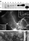
Figure 1
Figure 1
Specificity controls of the affinity-purified anti-PV1 rabbit IgGs. A, Immunoblotting of total membrane proteins (all lanes from one same gel) from cerebral cortex (Ctx, 100 mg), vmARC/ME (ME, 6 mg), whole pituitary (Pit, 20 mg), gastric antrum (Ga, 15 mg), duodenum (Du, 30 mg), uterus (Ut, 3 mg), adrenal gland (Ad, 20 mg), kidney (Kid, 20 mg), and lung (L, each 20 mg). Molecular masses (kDa) of markers (Bio-Rad, Marnets-la-Coquette, France) are on the left. Note the position of the ME band and that deglycosylation pretreatment (degly) reveals an apparent mass of 50 kDa consistent with the 50.04 kDa deduced from the open reading frame of the mRNA sequence. B, PV1 labeling over endothelial cells in the external zone of the ME (arrow in upper inset). The lower inset shows disk-shaped labeled structures (thin arrows; digitally enlarged from main view taken with a ×100 oil-immersion objective). Bar, 1 μm. C, Electron micrographs of two ultrathin sections (left, tangential; right, transverse) through external ME capillaries exhibiting immunogold (18 nm) labeling for PV1 at the central knob of fenestral diaphragms (arrows, discernible knobs without overlying immunogold). lu, Capillary lumen; pe, perivascular space. Bars, 100 nm.
Figure 2
Figure 2
Overview of PV1 mRNA expression in the brain of a male rat. PV1 mRNA expression is restricted to the CVOs and the pineal gland (thick arrows). Autoradiographic films after exposure to frontal sections hybridized with antisense or sense cRNA probes. Choroid plexi (ChP) are indicated by thin arrows. AP, area postrema; OVLT, organum vasculosum laminae terminalis; SFO, subfornical organ.
Figure 3
Figure 3
Overview of PV1-ir in brain of a male rat. PV1-ir decorates a majority of blood vessels in the CVOs and the pineal. Frontal sections double-labeled for PV1 (green) and the panendothelial cell marker RECA1 (red). Single optical sections (1.52 μm thick) from confocal microscopy. Note that blood vessels outside CVOs are PV1-negative and that all PV1-ir structures are also RECA1-positive. See Fig. 2 for legends. Bars, 50 μm.
Figure 4
Figure 4
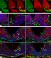
Permeable microvessels of the vmARC are bordered by chemosensory neurons and sexually dimorphic axons. A, PV1-labeling decorates all subsets of the vasculature of the vmARC/ME. Two adjacent frontal sections (50 μm thick; ∼−3.4 mm from bregma) double-labeled for PV1 (green) and either (red) the panendothelial cell marker RECA1 or the neuronal marker HuC/D (in merged images, PV1-signal is contrasted for clarity). In the vmARC (vm) numerous HuC/D-ir perikarya are close to PV1-ir subependymal capillaries (right, short arrows). icl, Intrainfundibular capillary loop; psp, primary superficial plexus; pv, long portal vessel; sep, subependymal plexus. *, Third ventricle. Bar, 100 μm. Normal male rat. B, Arcuate NPY/orexigenic and αMSH/anorexigenic sensor neurons lie in the vicinity of PV1-ir capillaries. Two frontal cuts at mid- (left) and retroinfundibular (right; ∼−3.8 mm from bregma) levels after triple-labeling (PV1, green; αMSH, blue; NPY, red) with high magnification views of boxed areas. Arrows point to peptidergic perikarya and arrowheads to putative reciprocal contacts. Optical sections from confocal microscopy (high powers are 0.38 μm thick). PS, Pituitary stalk; rME, retroinfundibular ME. Bars, 100 μm (low-power views) and 10 μm (high-power views). Two colchicine-treated females. C, Permeable PV1-ir (green, arrows) microvessels have similar distribution in the vmARC and rME in both sexes but are bordered by palisades of NKB-ir axons (blue,arrowheads; purposely lightened for contrast) in males only. Red labeling is either for HuC/D or RECA1, as indicated. Single 1.52-μm-thick optical cuts of triple-labeled, adjacent cryostat sections (∼−3.8 mm from bregma) from normal rats. Bar, 50 μm.
Figure 5
Figure 5
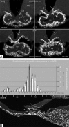
Topographic distribution of PV1-ir capillaries in the vmARC. A, The highly permeable portion of the vmARC proximal to the pituitary stalk (PS). Labeling for PV1 in consecutive frontal sections (50 μm thick) revealing how sinusoidal capillaries branch up from the primary superficial plexus (arrow in 2) to give off ramifications of the subependymal plexus (other arrows) in the vmARC (vm). Labeling is variable among capillaries. Female rat at 1800 h on proestrus. *, Infundibular recess of the third ventricle. Bar, 100 μm. B (Upper panel), Topographical analysis in male rats (n = 12) of the number (mean ± sem) of capillary profiles (RECA1-ir) with PV1-labeling (white bars) and numbers of PV1-ir pixels (gray bars) after bilateral sampling of the vmARC in coronal 50-μm-thick sections regularly spaced by 150 μm. Permeable microvessels were more frequently observed proximal to the pituitary stalk (planes 11–14). Most section planes significantly differed from one another by ANOVA, which is not indicated for purpose of clarity. B (Bottom panel), Cryostat-cut parasagittal section of a male rat hypothalamus after labeling for PV1 showing a permeable sinusoid (arrow) entering the retroinfundibular vmARC (vm) about −3.9 mm from bregma at section plane 14.
Figure 6
Figure 6
Analysis of the distribution of PV1-labeling in the vmARC from section planes 1-16 in adult male and female rats (data are given as mean ± sem numbers of PV1-ir capillary profiles and PV-ir pixels counted bilaterally; see Materials and Methods, supplemental Methods and supplemental Table 1). A, Comparison of males (n = 12) with females killed at 1000 h the day of diestrus 1 (D1; n = 10), or the day of proestrus at 1000 h (P10; n = 7) or 2000 h (P20; n = 9). B, Height groups of proestrous females killed every 2 h from 0800–2200 h (n = 4–6/time point). Statistical analysis was performed with groups either taken individually (B1) or grouped (B2) according to animals’ low presurge (n = 23) or high surge (n = 14) LH levels (see supplemental Table 1). The two-way repeated measure ANOVA detected an effect of topography for all group comparisons, but no effect of sex/cycle (A) or time of the day (B1). In B2, there was, in addition by post hoc Tukey’s multiple comparison test, an interaction (topography × LH) visible at planes 11 and 12 (**, P < .01). In A, B1, and B2, most section planes significantly differed from one another, which is not indicated for purpose of clarity.
Figure 7
Figure 7
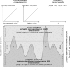
Hypothesis of a biphasic action of circulating hormones upon the arcuate nucleus. Top, The role of the feedback sensor systems of the ARC is to ensure reliability of hypothalamic (hypo.) output in the face of peripheral (periph. resp.) sensitivity to pituitary action, i.e. in a context-specific manner. t, Time. Bottom, The sensor systems of the vmARC may be subjected to physiological regulation of their sensitivity (threshold) to both amplitude and frequency of feedback signal, e.g. as exemplified here, may be able to either see two, three, or five feedback pulses (numbers in same row indicate how many pulses would be detected at three different levels of sensitivity). See Discussion.

References

    1. Grove KL, Grayson BE, Glavas MM, Xiao XQ, Smith MS 2005 Development of metabolic systems. Physiol Behav 86:646–660 - PubMed
    1. Cone RD 2006 Studies on the physiological functions of the melanocortin system. Endocr Rev 27:736–749 - PubMed
    1. Morton GJ, Cummings DE, Baskin DG, Barsh GS, Schwartz MW 2006 Central nervous system control of food intake and body weight. Nature 443:289–295 - PubMed
    1. Plum L, Belgardt BF, Brüning JC 2006 Central insulin action in energy and glucose homeostasis. J Clin Invest 116:1761–1766 - PMC - PubMed
    1. Badman MK, Flier JS 2007 The adipocyte as an active participant in energy balance and metabolism. Gastroenterology 132:2103–2115 - PubMed

Publication types

MeSH terms