Surgical removal of right-to-left cardiac shunt in the American alligator (Alligator mississippiensis) causes ventricular enlargement but does not alter apnoea or metabolism during diving
- PMID: 19837897
- PMCID: PMC3188805
- DOI: 10.1242/jeb.034595
Surgical removal of right-to-left cardiac shunt in the American alligator (Alligator mississippiensis) causes ventricular enlargement but does not alter apnoea or metabolism during diving
Abstract
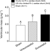
Crocodilians have complete anatomical separation between the ventricles, similar to birds and mammals, but retain the dual aortic arch system found in all non-avian reptiles. This cardiac anatomy allows surgical modification that prevents right-to-left (R-L) cardiac shunt. A R-L shunt is a bypass of the pulmonary circulation and recirculation of oxygen-poor blood back to the systemic circulation and has often been observed during the frequent apnoeic periods of non-avian reptiles, particularly during diving in aquatic species. We eliminated R-L shunt in American alligators (Alligator mississippiensis) by surgically occluding the left aorta (LAo; arising from right ventricle) upstream and downstream of the foramen of Panizza (FoP), and we tested the hypotheses that this removal of R-L shunt would cause afterload-induced cardiac remodelling and adversely affect diving performance. Occlusion of the LAo both upstream and downstream of the FoP for approximately 21 months caused a doubling of RV pressure and significant ventricular enlargement (average approximately 65%) compared with age-matched, sham-operated animals. In a separate group of recovered, surgically altered alligators allowed to dive freely in a dive chamber at 23 degrees C, occlusion of the LAo did not alter oxygen consumption or voluntary apnoeic periods relative to sham animals. While surgical removal of R-L shunt causes considerable changes in cardiac morphology similar to aortic banding in mammals, its removal does not affect the respiratory pattern or metabolism of alligators. It appears probable that the low metabolic rate of reptiles, rather than pulmonary circulatory bypass, allows for normal aerobic dives.
Figures


Similar articles
-
Role of the left aortic arch and blood flows in embryonic American alligator (Alligator mississippiensis).J Comp Physiol B. 2011 Apr;181(3):391-401. doi: 10.1007/s00360-010-0494-6. Epub 2010 Oct 30. J Comp Physiol B. 2011. PMID: 21053004 Free PMC article.
-
Turning crocodilian hearts into bird hearts: growth rates are similar for alligators with and without right-to-left cardiac shunt.J Exp Biol. 2010 Aug 1;213(Pt 15):2673-80. doi: 10.1242/jeb.042051. J Exp Biol. 2010. PMID: 20639429 Free PMC article.
-
Effects of prolonged lung inflation or deflation on pulmonary stretch receptor discharge in the alligator (Alligator mississippiensis).Respir Physiol Neurobiol. 2014 Aug 15;200:25-32. doi: 10.1016/j.resp.2014.05.006. Epub 2014 May 27. Respir Physiol Neurobiol. 2014. PMID: 24874556
-
The crocodilian heart and central hemodynamics.Cardioscience. 1994 Sep;5(3):163-6. Cardioscience. 1994. PMID: 7827252 Review.
-
Elucidating the responses and role of the cardiovascular system in crocodilians during diving: fifty years on from the work of C.G. Wilber.Comp Biochem Physiol A Mol Integr Physiol. 2011 Sep;160(1):1-8. doi: 10.1016/j.cbpa.2011.05.015. Epub 2011 May 25. Comp Biochem Physiol A Mol Integr Physiol. 2011. PMID: 21635959 Review.
Cited by
-
Vagal tone regulates cardiac shunts during activity and at low temperatures in the South American rattlesnake, Crotalus durissus.J Comp Physiol B. 2016 Dec;186(8):1059-1066. doi: 10.1007/s00360-016-1008-y. Epub 2016 Jun 13. J Comp Physiol B. 2016. PMID: 27294346
-
Hemodynamics During Development and Postnatal Life.Adv Exp Med Biol. 2024;1441:201-226. doi: 10.1007/978-3-031-44087-8_11. Adv Exp Med Biol. 2024. PMID: 38884713 Review.
-
Role of the left aortic arch and blood flows in embryonic American alligator (Alligator mississippiensis).J Comp Physiol B. 2011 Apr;181(3):391-401. doi: 10.1007/s00360-010-0494-6. Epub 2010 Oct 30. J Comp Physiol B. 2011. PMID: 21053004 Free PMC article.
-
Turning crocodilian hearts into bird hearts: growth rates are similar for alligators with and without right-to-left cardiac shunt.J Exp Biol. 2010 Aug 1;213(Pt 15):2673-80. doi: 10.1242/jeb.042051. J Exp Biol. 2010. PMID: 20639429 Free PMC article.
-
Defibrillate You Later, Alligator: Q10 Scaling and Refractoriness Keeps Alligators from Fibrillation.Integr Org Biol. 2021 Jan 27;3(1):obaa047. doi: 10.1093/iob/obaa047. eCollection 2021. Integr Org Biol. 2021. PMID: 33977229 Free PMC article.
References
-
- Axelsson, M. (2001). The crocodilian heart; more controlled than we thought? Exp. Physiol. 86,785 -789. - PubMed
-
- Axelsson, M. and Franklin, C. E. (2001). The calibre of the Foramen of Panizza in Crocodylus porosus is variable and under adrenergic control. J. Comp. Physiol. B. 171,341 -346. - PubMed
-
- Axelsson, M., Holm, S. and Nilsson, S. (1989). Flow dynamics of the Crocodilian heart. Am. J. Physiol. 256,R875 -R879. - PubMed
-
- Axelsson, M., Fritsche, R., Holmgren, S., Grove, D. J. and Nilsson, S. (1991). Gut blood flow in the estuarine crocodile, Crocodylus porosus. Acta. Physiol. Scand. 142,509 -516. - PubMed
-
- Axelsson, M., Franklin, C. E., Lofman, C. O., Nilsson, S. and Grigg, G. (1996). Dynamic anatomical study of cardiac shunting in crocodiles using high-resolution angioscopy. J. Exp. Biol. 199,359 -365. - PubMed
Publication types
MeSH terms
Grants and funding
LinkOut - more resources
Full Text Sources

