. 2010 Feb;9(2):197-208.
doi: 10.1074/mcp.M900152-MCP200. Epub 2009 Oct 16.

Proteomics analysis of A33 immunoaffinity-purified exosomes released from the human colon tumor cell line LIM1215 reveals a tissue-specific protein signature

Affiliations

Proteomics analysis of A33 immunoaffinity-purified exosomes released from the human colon tumor cell line LIM1215 reveals a tissue-specific protein signature

Suresh Mathivanan et al. Mol Cell Proteomics. 2010 Feb.

Abstract

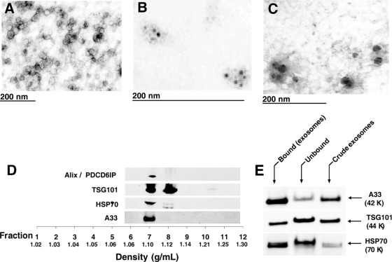
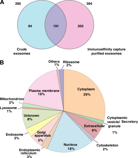
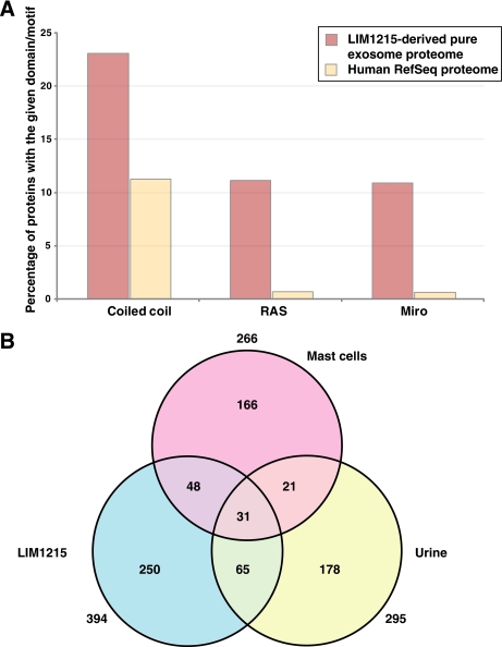
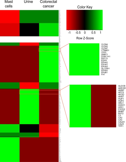
Exosomes are 40-100-nm-diameter nanovesicles of endocytic origin that are released from diverse cell types. To better understand the biological role of exosomes and to avoid confounding data arising from proteinaceous contaminants, it is important to work with highly purified material. Here, we describe an immunoaffinity capture method using the colon epithelial cell-specific A33 antibody to purify colorectal cancer cell (LIM1215)-derived exosomes. LC-MS/MS revealed 394 unique exosomal proteins of which 112 proteins (28%) contained signal peptides and a significant enrichment of proteins containing coiled coil, RAS, and MIRO domains. A comparative protein profiling analysis of LIM1215-, murine mast cell-, and human urine-derived exosomes revealed a subset of proteins common to all exosomes such as endosomal sorting complex required for transport (ESCRT) proteins, tetraspanins, signaling, trafficking, and cytoskeletal proteins. A conspicuous finding of this comparative analysis was the presence of host cell-specific (LIM1215 exosome) proteins such as A33, cadherin-17, carcinoembryonic antigen, epithelial cell surface antigen (EpCAM), proliferating cell nuclear antigen, epidermal growth factor receptor, mucin 13, misshapen-like kinase 1, keratin 18, mitogen-activated protein kinase 4, claudins (1, 3, and 7), centrosomal protein 55 kDa, and ephrin-B1 and -B2. Furthermore, we report the presence of the enzyme phospholipid scramblase implicated in transbilayer lipid distribution membrane remodeling. The LIM1215-specific exosomal proteins identified in this study may provide insights into colon cancer biology and potential diagnostic biomarkers.


Figures

Fig. 1.
Fig. 1.
Morphological characterization and proteomics analysis of LIM1215 colorectal cancer-derived exosomes. Electron micrographs of crude exosomes negatively stained with uranyl acetate and examined at 60 kV (A), anti-A33 (B), and anti-CD63 immunogold/uranyl acetate staining (C) are shown. D, OptiPrep density gradient separation of crude exosomes using Alix/PDCD6IP, TSG101, HSP70, and A33 as exosomal markers to locate the fraction of exosome sedimentation. E, Western blot analysis of bound, unbound, and crude exosomes (5 μg/lane) for A33 antigen, TSG101, and HSP70.
Fig. 2.
Fig. 2.
Overlap of crude and immunoaffinity capture-purified exosomal proteomes and subcellular localization distribution of LIM1215-derived pure exosomal proteins. A, Venn diagram depicting the overlap of exosomal proteomes. The numbers present outside the circle represent the total number of proteins identified in the particular data set. A two-way Venn diagram of the LIM1215 exosome proteome between crude exosomes and the immunoaffinity capture-based purified data set is depicted. 191 proteins are common between the two exosomal data sets. 394 proteins found in the immunoaffinity capture-based purified exosomes were used in further analysis. B, subcellular localization of the 394 proteins identified in the LIM1215-derived pure exosomes is shown. 29% of the proteins have a cytoplasmic localization, whereas 19% of the proteins have plasma membrane localization. 6% of the proteins are known to be extracellular, and 20% are known to be localized to the nucleus.
Fig. 3.
Fig. 3.
Protein domain distribution of LIM1215-derived pure exosomal proteins and overlap of urine-, mast cell-, and LIM1215-derived pure exosomal proteomes. A, distribution of protein domains that are preferentially enriched in the LIM1215-derived pure exosomes. Coiled coil domain is predicted to be present in ∼23% of the exosomal proteome, whereas it is present in only ∼11% of the entire human proteome (human RefSeq proteome). Likewise, RAS and MIRO domains are highly significant in the LIM1215-derived pure exosomal proteomes (11%) as compared with the entire human proteome (0.7%). B, three-way Venn diagram depicting the overlap between the exosomal proteomes derived from urine, mast cells, and LIM1215 cells. Here, two previous exosomal studies published in the scientific literature were used to find the overlap between the exosomal proteomes. 31 proteins were found to be identified in all three exosomal proteomes, whereas 96 and 79 proteins were found to be in common between LIM1215-urine and LIM1215-mast cell exosome data sets, respectively.
Fig. 4.
Fig. 4.
Tissue-specific signature of exosomal proteins. A heat map of exosomal proteomes along with the protein tissue expression is shown. Hierarchical clustering of the matrix plotted by the presence or absence of a specific protein in a particular exosomal data set and its corresponding protein tissue expression profiles shows proteins that are expressed in a particular tissue type as compared with the others. Deep red indicates that the protein is not known to be found in the specific tissue and not identified in the corresponding exosomal data set as well. Bright green indicates that the protein is identified in the exosomal data set and also in the specific tissue. Protein tissue profiles were downloaded from Human Proteinpedia, HPRD, Human Protein Atlas, and the urinary proteome data set. The top zoom panel displays a subset of proteins that are uniquely identified in the LIM1215 (colorectal cancer)-derived pure exosomes and are also known to be expressed in colorectal cancer tissues. The bottom zoom panel shows subset of proteins that are uniquely identified in urine-derived exosomes.

References

    1. Simpson R. J., Jensen S. S., Lim J. W. ( 2008) Proteomic profiling of exosomes: current perspectives. Proteomics 8, 4083– 4099 - PubMed
    1. Pisitkun T., Shen R. F., Knepper M. A. ( 2004) Identification and proteomic profiling of exosomes in human urine. Proc. Natl. Acad. Sci. U.S.A 101, 13368– 13373 - PMC - PubMed
    1. Taylor D. D., Akyol S., Gercel-Taylor C. ( 2006) Pregnancy-associated exosomes and their modulation of T cell signaling. J. Immunol 176, 1534– 1542 - PubMed
    1. Sabapatha A., Gercel-Taylor C., Taylor D. D. ( 2006) Specific isolation of placenta-derived exosomes from the circulation of pregnant women and their immunoregulatory consequences. Am. J. Reprod. Immunol 56, 345– 355 - PubMed
    1. Andre F., Schartz N. E., Movassagh M., Flament C., Pautier P., Morice P., Pomel C., Lhomme C., Escudier B., Le Chevalier T., Tursz T., Amigorena S., Raposo G., Angevin E., Zitvogel L. ( 2002) Malignant effusions and immunogenic tumour-derived exosomes. Lancet 360, 295– 305 - PubMed

Publication types