. 2009 Nov;12(11):1398-406.
doi: 10.1038/nn.2410. Epub 2009 Oct 18.

The oligodendrocyte-specific G protein-coupled receptor GPR17 is a cell-intrinsic timer of myelination

Affiliations

The oligodendrocyte-specific G protein-coupled receptor GPR17 is a cell-intrinsic timer of myelination

Ying Chen et al. Nat Neurosci. 2009 Nov.

Abstract

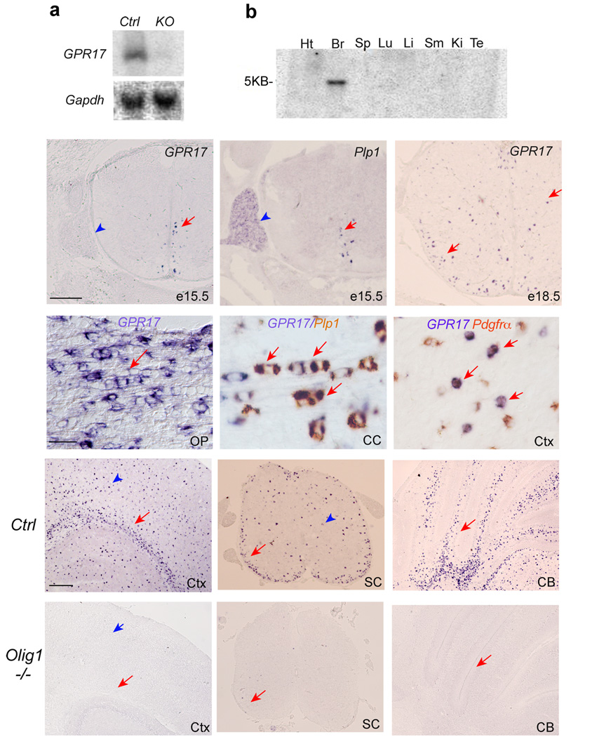
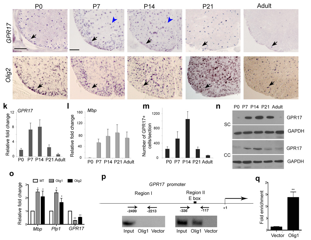
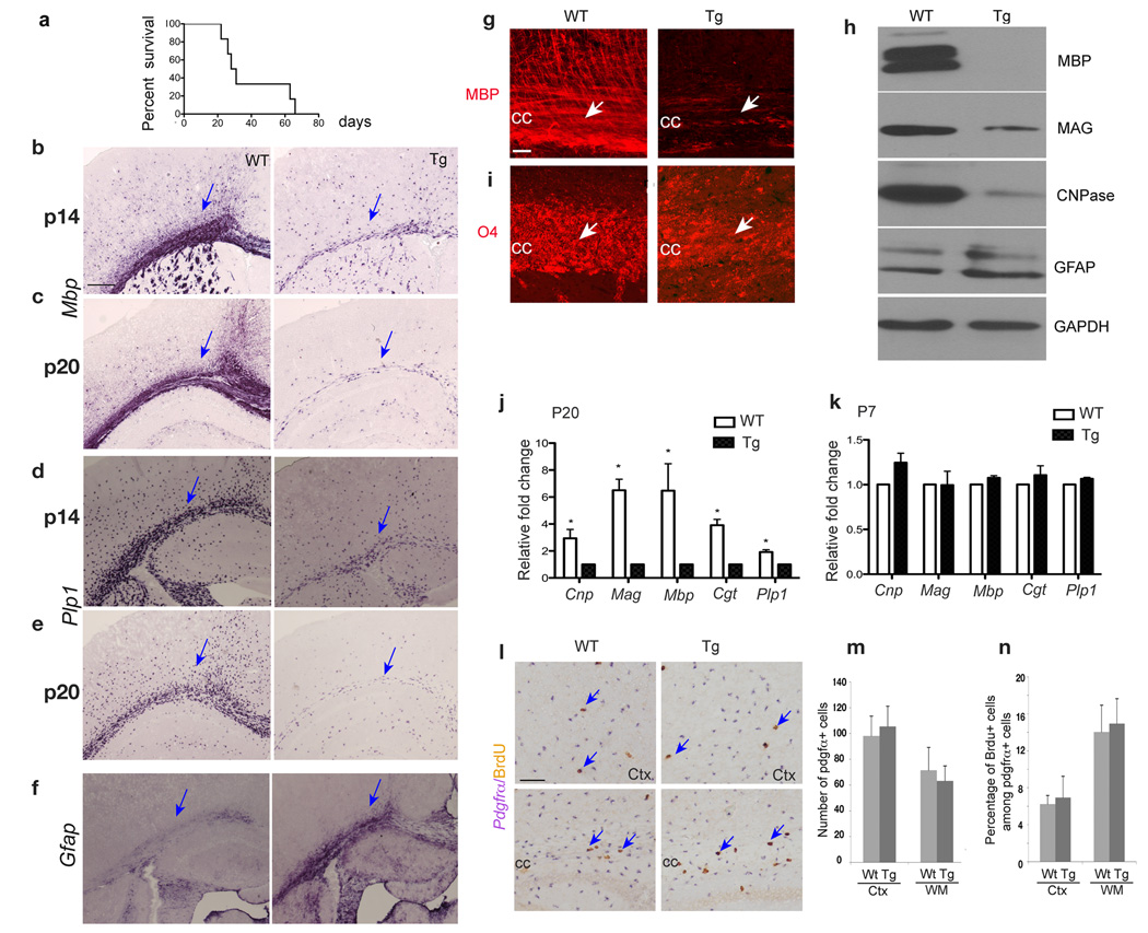
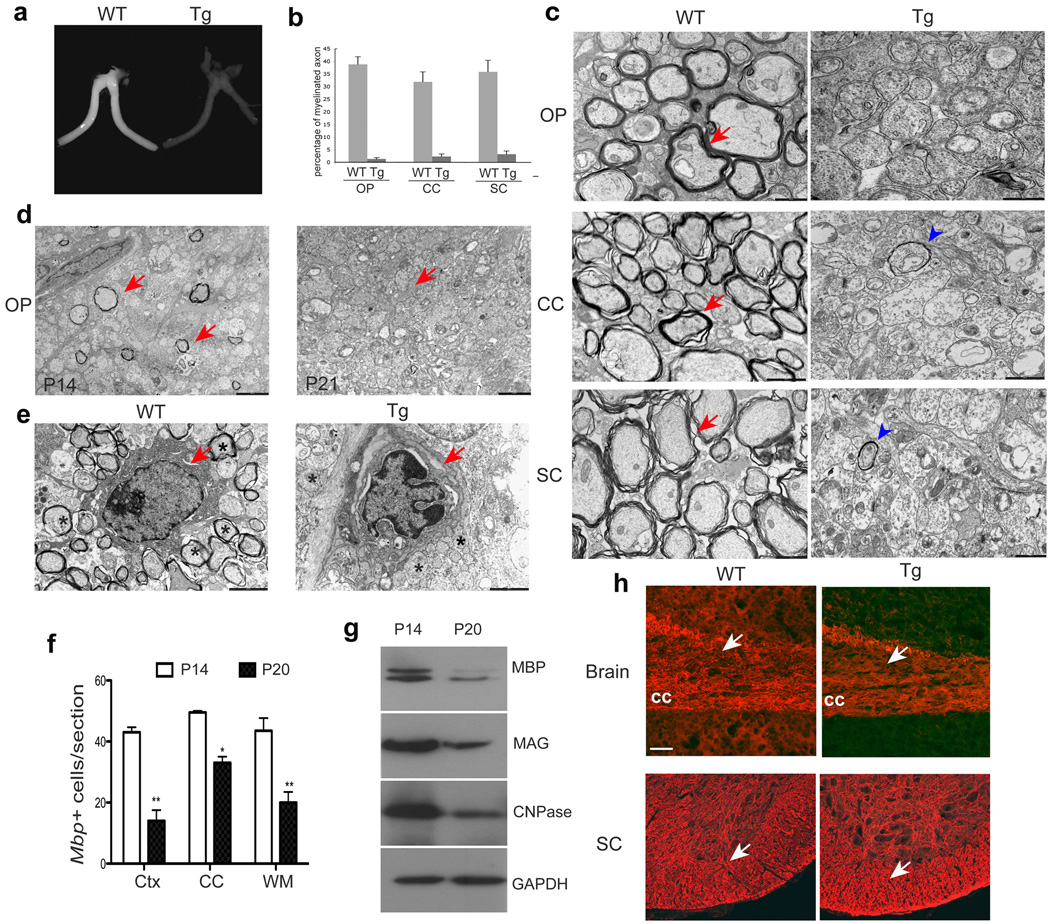
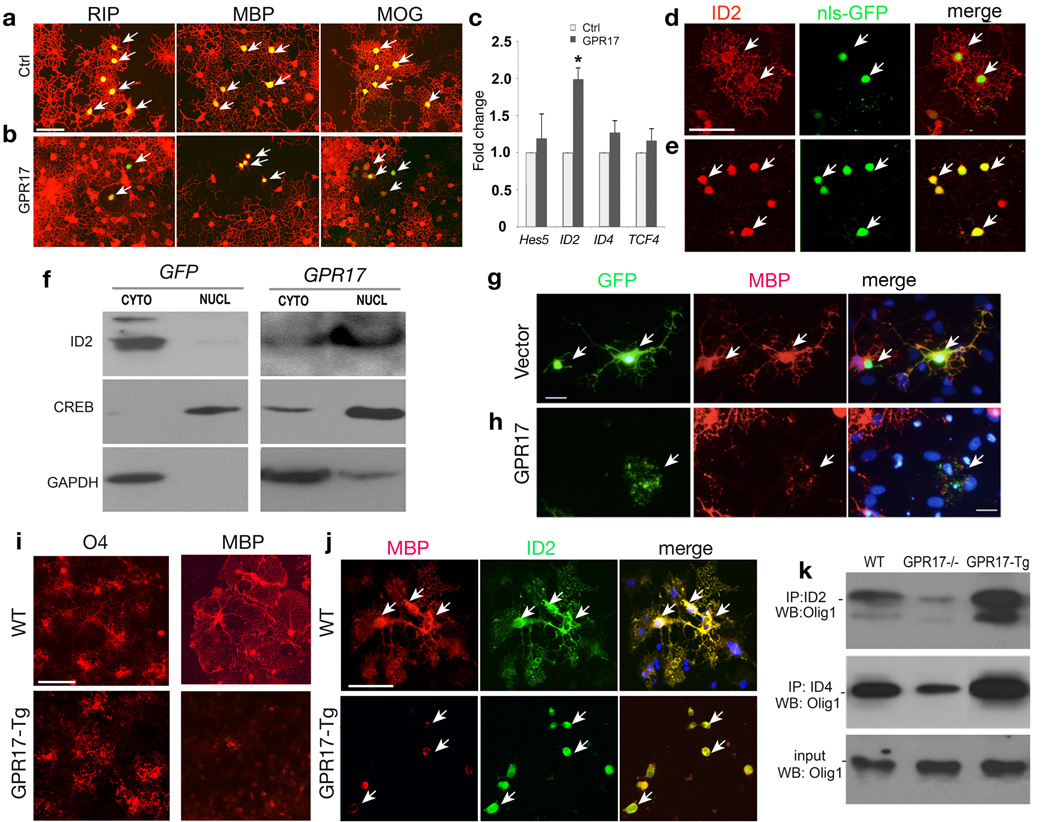
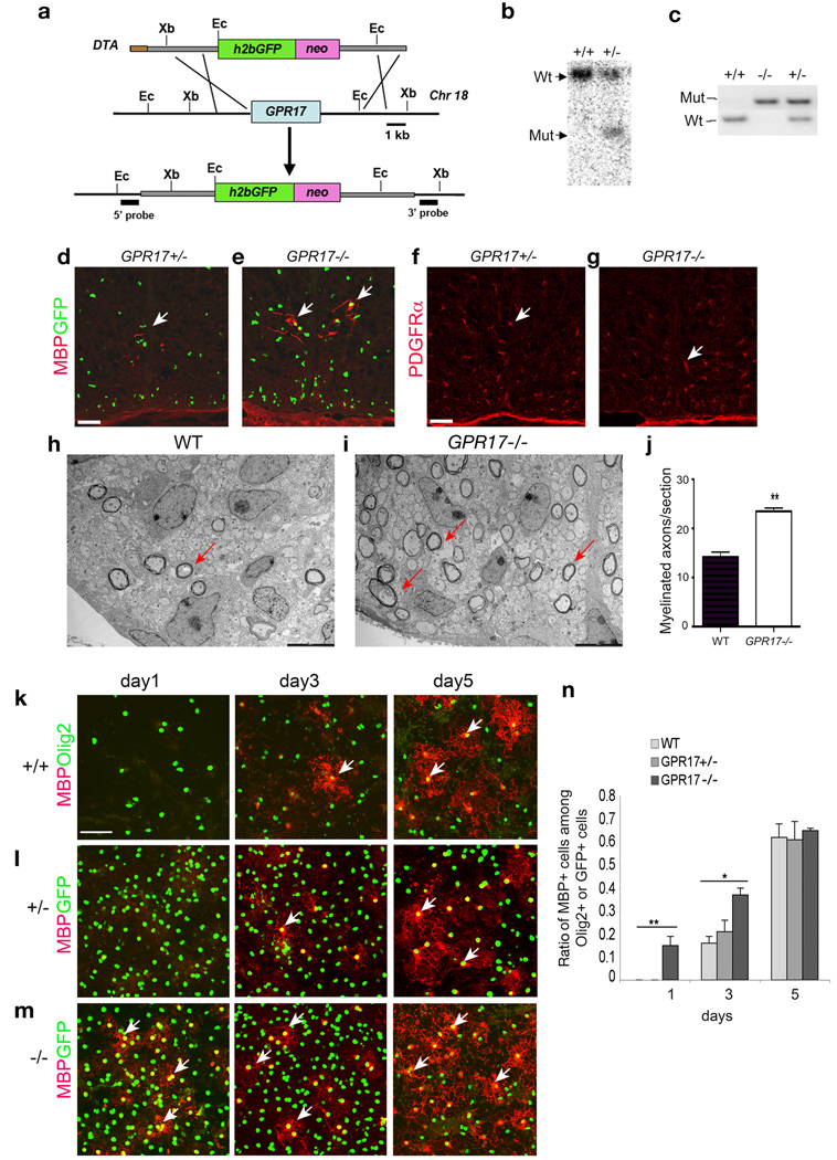
The basic helix-loop-helix transcription factor Olig1 promotes oligodendrocyte maturation and is required for myelin repair. We characterized an Olig1-regulated G protein-coupled receptor, GPR17, whose function is to oppose the action of Olig1. Gpr17 was restricted to oligodendrocyte lineage cells, but was downregulated during the peak period of myelination and in adulthood. Transgenic mice with sustained Gpr17 expression in oligodendrocytes exhibited stereotypic features of myelinating disorders in the CNS. Gpr17 overexpression inhibited oligodendrocyte differentiation and maturation both in vivo and in vitro. Conversely, Gpr17 knockout mice showed early onset of oligodendrocyte myelination. The opposing action of Gpr17 on oligodendrocyte maturation reflects, at least partially, upregulation and nuclear translocation of the potent oligodendrocyte differentiation inhibitors ID2/4. Collectively, these findings suggest that GPR17 orchestrates the transition between immature and myelinating oligodendrocytes via an ID protein-mediated negative regulation and may serve as a potential therapeutic target for CNS myelin repair.


Figures

Figure 1
Figure 1. Identification and expression of GPR17 in the murine CNS
a) Upper panel, a Northern blot of RNA extracted from brain tissues of WT and Olig1 mutant mice at P14 was probed with 32P-labeled GPR17. Lower panel, expression of a housekeeping gene Gapdh from the same samples above was used as loading controls. b) A commercial Northern blot (Clonetech) of mRNA extracted from various rat tissues was probed with 32P-labeled GPR17, revealing 5kb mRNA transcript in the brain. c–f) In situ hybridization was performed with probes specific for GPR17 and Plp1/DM20 in e15.5 (c–d) and e18.5 (e) spinal cords and P14 (f) optic nerve in mice. Arrows indicate labeled cells. Arrowheads indicate dorsal root ganglia. g–h) Double in situ hybridization labeling for GPR17 (blue color) and Plp1 (brown color) or Pdgfrα (brown color) at P14. Arrows indicate a linear array of interfascicular oligodendrocytes expressing both GPR17 and Plp1 in the corpus callosum (g). Arrows in h indicate GPR17 expression in a subset of PDGFRα+ OPCs in the cerebral cortex. i–n) Expression of GPR17 mRNA in various CNS regions of WT (i–k) and Olig1KO (l–n) mice at P14. Arrows and arrowheads indicate labeling cells in white matter tracts and grey matter regions, respectively. SC, spinal cord; CB, cerebellum; OP, optic nerve; CC, corpus callosum; Ctx, cortex. Scale bars in c–e: 200 µm; f–h, 100 µm; i–n: 200 µm.
Figure 2
Figure 2. Transient expression of GPR17 in oligodendrocytes during development
a–j) In situ hybridization on transverse spinal cord sections from P0, P7, P14, P21 and adulthood with probes to murine GPR17 and Olig2 as indicated. Arrows and arrowheads indicate GPR17 or Olig2 positive cells in the spinal white or gray matter, respectively. k–l) Transcripts of GPR17 (k) and Mbp (l) were determined in total RNA prepared from spinal cords at different stages by qRT-PCR (n=3). m) Average number of GPR17+ cells per spinal section at the indicated age. GPR17+ cell number was counted from at least 5 sections at the thoracic level of each animal (n=3). n) Western blot analysis of GPR17 protein from the spinal cord and corpus callosum at indicated stages. GAPDH was used as a loading control for protein extracts. o) qRT-PCR analysis of GPR17, Mbp and Plp1 transcript in adult hippocampus-derived neural stem/progenitor cells (HCN) transfected with pCS2MT-nls-Olig1, Olig2 or control expression vector. The fold change of gene expression was present from cells transfected with Olig1 or Olig2 versus control (*P<0.01, **P<0.001, Student t-test). p) Recruitment of Olig1 to the GPR17 5’ upstream regulatory sequence. Regions upstream the start site of GPR17 with E-box (Region II) or without E-Box (Region I) consensus sequences were amplified with specific PCR primers for ChIP assay. Input DNA was used as positive control. q) Quantification of ChIP enrichment of Olig1 binding to E-box containing region II in the GPR17 promoter by qRT-PCR in Olig1-transfected cells compared to vector-transfected cells (**P<0.001, Student t-test). Scale bars in a–j: 200 µm.
Figure 3
Figure 3. Deficiency of myelin gene expression in the brain of GPR17 transgenic animals
a) Life span assessment shows the majority of CNP1-GPR17 transgenic mice die around postnatal 3–4 weeks (17/23 examined). A few (6/23) transgenic mice can barely survive to the adulthood. b–f) In situ hybridization was performed with antisense riboprobes against Mbp, Plp1 and Gfap on the forebrain of WT and GPR17 transgenic (Tg) littermates at P14 and P20. Arrows indicate the white matter. g, i) Immunohistochemistry was performed with antibodies against MBP and O4 on the corpus callosum (CC, arrows) sections from WT and Tg littermates at P20. h) Western blot analysis of MBP, MAG, CNPase and GFAP on the tissues from the forebrain of WT and Tg littermates at P19. j–k) Expression of Cnp, Mag, Mbp, Cgt and Plp1 was determined using total RNAs prepared from the forebrain of WT and Tg littermates at P7 and P20 by qRT-PCR from three independent experiments. Values were normalized to GAPDH for each sample. *P<0.01 (Student t-test). l) Cycling OPCs in the brain were determined by double in situ hybridization and immunohistochemistry 4 h after BrdU administration to animals at P14. BrdU+ (brown)/Pdgfrα+ (purple) OPCs are indicated by arrows. m, n) The number of Pdgfrα+ OPCs per field (0.08 mm2) (m) and the proportion of BrdU+/Pdgfrα+ OPCs in the cell cycle (n) were quantified from three WT and Tg littermates. cc, corpus callosum; Ctx, cortex. Scale bars: b–f, g, i and l, 100 µm.
Figure 4
Figure 4. Myelinogenesis defects in the CNS of CNP-GPR17 transgenic mice
a) Optic nerves isolated from WT mice at P19. b) Quantification of the proportion of myelinating axons from the optic nerve, corpus callosum and spinal cord at P19 (>800 axons scored). c) Electron micrographs of the optic nerve, corpus callosum and spinal cord in cross-sections from WT and Tg mice at P19. Arrows and arrowheads indicate myelin sheaths. d) Electron micrographs of cross-sections of optic nerves from Tg mice at P14 and P21. Arrows indicate myelinated axons, which are absent in the optic nerve of P21 Tg animals. e) Ultrastructural analysis showing an apoptotic oligodendrocyte in optic nerves of Tg mice, but a healthy oligodendrocyte in WT mice at P16 (Arrows). Asterisks indicate myelinated axons in WT and unmyelinated axons in Tg mice. f) Mbp expression on brain sections of Tg mice at P14 and P20 was detected by in situ hybridization. The number of Mbp+ oligodendrocytes per field (0.08 mm2) at corresponding regions was quantified in the corpus callosum (CC), cortex (Ctx), and white matter (WM). *P<0.01, **P<0.001 (Student t-test, n=3). g) Western blot analysis of MBP, MAG, CNPase from the forebrain of Tg mice at P14 and P20. GAPDH as a control. h) The sections from the brain and spinal cord of WT and Tg mice at P20 were immuostained with Neurofilament (red). Arrows indicate the corpus callosum and spinal white matter, respectively. Scale bars in c–e, 1 µm; h: 50 µm.
Figure 5
Figure 5. GPR17 overexpression inhibits oligodendrocyte differentiation and induces nuclear translocation of ID2/4
a–b) HCN cells were transfected with GPR17 and control vector carrying GFP and assayed after 72 hrs and immunostained for RIP, MBP and MOG. Arrows indicate the transfected cells. c) Total RNAs extracted from HCN cells transfected with control and GPR17 were subjected to qRT-PCR analysis for Hes5, ID2, ID4 and TCF4. *P<0.01 (Student t-test). d–e) HCN cells transfected with GPR17 (e) or control pCIG (d) carrying nuclear-localized GFP (nls-GFP) were immunostained for ID2. Arrows indicate ID2 immunoreactivity in control- or GPR17-transfected cells, respectively. f) Cytoplasmic (CYTO) and nuclear (NUCL) fractions were prepared 3 days after transfection and subjected to Western blotting analysis for cellular localization of ID2, CREB or GAPDH. g–h) Primary rat OPCs transfected with control or GPR17-expression vector were co-cultured with astrocytes for 5 days and subjected to immunocytochemistry for MBP and GFP. Arrows in g, h indicate MBP+ cells in control- or GPR17-transfected cells. i–j) Cortical progenitors from WT and GPR17-Tg embryos at E15.5 were cultured to promote oligodendrocyte formation. Cells were immunostained with antibodies to O4, MBP and ID2 as indicated. Arrows in j indicate MBP or ID2 expression. k) Cortical progenitor cells from WT, GPR17−/− and CNP1-GPR17 transgenic embryos at E15.5 were cultured to promote oligodendrocyte formation. The cells were collected and processed for co-immunoprecipitation with an antibody against ID2 or ID4. Immunoprecipitated proteins were subjected to Western blot analysis for Olig1 as indicated. Scale bars in a–e, 50 mm; g–h; 25 mm and i–j, 50 mm.
Figure 6
Figure 6. Early onset of oligodendrocyte myelination in GPR17 null mice
a) Schematic strategy for disruption of GPR17 on the mouse chromosome 18. Abbreviations: h2bGFP, histone2b GFP; neo, the neomycin selection marker; and DTA, the diphtheria toxin gene. Ec: EcoRI, Xb: XbaI. b) Southern blot validation with a 5′ GPR17 probe identified the targeted locus as a following EcoRI digestion of genomic DNAs. c) PCR genotyping analysis of WT and targeted allele from WT, GPR17+/− and −/− animals. d–g) Sections from the spinal cord of GPR17+/− and −/− embryos at E17.5 were immunostained for MBP (d, e) and PDGFRα (f, g). Arrows indicate MBP+ and PDGFRα+ cells, respectively. h–i) Electron micrographs of spinal cross-sections of WT and GPR17−/− mice at P3. Arrows indicate multilamellar myelin sheaths around axons in the lateral white matter. j) Quantitative analysis of myelinated axon fibers in GPR17−/− and WT mice at P3 in the corresponding region of lateral white matter of spinal cord (**P<0.001, Student t-test). k–m) Cortical progenitor cells from WT, GPR17+/− and −/− embryos at E15.5 were cultured to promote oligodendrocyte differentiation. Cells were then immunostained at defined days as indicated for MBP (arrows) and GFP (l–m) or Olig2 (k). n) Quantification of cells expressing MBP from above cultures. All data shown are derived from three experiments in parallel cultures of at least three age-matching littermates (> 1500 cell counts at each stage). Error bars represent SDs. **p<0.001, One-way ANOVA with Newman-Keuls Multiple comparison test. Scale bars: d–g, 100 µm; h–i, 1 µm and k–m, 50 µm.

Similar articles

Cited by

References

    1. Trapp BD, et al. Axonal transection in the lesions of multiple sclerosis. N Engl J Med. 1998;338:278–285. - PubMed
    1. Berger J, Moser HW, Forss-Petter S. Leukodystrophies: recent developments in genetics, molecular biology, pathogenesis and treatment. Curr Opin Neurol. 2001;14:305–312. - PubMed
    1. Pfeiffer SE, Warrington AE, Bansal r. The oligodendrocyte and its many cellular processes. Trends in Cell Biology. 1993;3:191–197. - PubMed
    1. Wang S, Sdrulla A, Johnson JE, Yokota Y, Barres BA. A role for the helix-loop-helix protein Id2 in the control of oligodendrocyte development. Neuron. 2001;29:603–614. - PubMed
    1. Kondo T, Raff M. The Id4 HLH protein and the timing of oligodendrocyte differentiation. Embo J. 2000;19:1998–2007. - PMC - PubMed

MeSH terms

Substances