. 2010 Jan;9(1):82-8.
doi: 10.1038/nmat2563. Epub 2009 Oct 18.

Material properties of the cell dictate stress-induced spreading and differentiation in embryonic stem cells

Affiliations

Material properties of the cell dictate stress-induced spreading and differentiation in embryonic stem cells

Farhan Chowdhury et al. Nat Mater. 2010 Jan.

Abstract

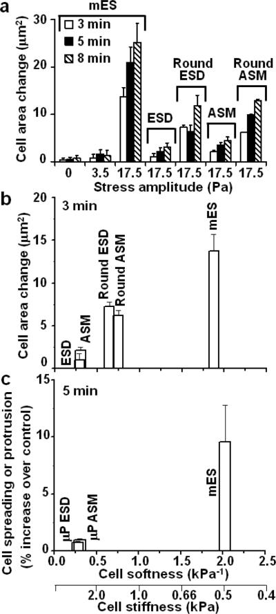
Growing evidence suggests that physical microenvironments and mechanical stresses, in addition to soluble factors, help direct mesenchymal-stem-cell fate. However, biological responses to a local force in embryonic stem cells remain elusive. Here we show that a local cyclic stress through focal adhesions induced spreading in mouse embryonic stem cells but not in mouse embryonic stem-cell-differentiated cells, which were ten times stiffer. This response was dictated by the cell material property (cell softness), suggesting that a threshold cell deformation is the key setpoint for triggering spreading responses. Traction quantification and pharmacological or shRNA intervention revealed that myosin II contractility, F-actin, Src or cdc42 were essential in the spreading response. The applied stress led to oct3/4 gene downregulation in mES cells. Our findings demonstrate that cell softness dictates cellular sensitivity to force, suggesting that local small forces might have far more important roles in early development of soft embryos than previously appreciated.


Figures

Figure 1
Figure 1. Cell softness dictates cell spreading response to stress
a, Stress-induced spreading in mES cells is amplitude-dependent. Amplitude is the magnitude of change in a sinusoidal oscillatory forcing system where the mean magnitude is zero. ES cells did not spread at 0 or 3.5-Pa stress but started to protrude and spread at 17.5-Pa stress (n=7, 5, or 9 cells for 0, 3.5, or 17.5 Pa stress, respectively). There were no significant differences in cell area change between 0 and 3.5-Pa stress (p>0.58, 0.23, or 0.68 at 3, 5, or 8 min). In contrast, there were significant differences between 3.5 and 17.5 Pa stress (p<0.0007, 4.92×10⁻⁵, or 5.66×10⁻⁵ at 3, 5 or 8 min respectively). At 17.5 Pa stress, there was significant difference in cell area between 3 min and 5 min (p<0.05), but no significant difference in cell area between 5 min and 8 min (p>0.23). In sharp contrast, for ESD cells and ASM cells there were no stress-induced changes in cell area even at 17.5-Pa applied stress (n= 7 cells for both cell types). There were no significant differences in cell area change between 3 min and 5 min (p>0.30 for ESD and p>0.09 for ASM) or 5 min and 8 min (p>0.47 for ESD and p>0.37 for ASM). Round ESD cells and round ASM cells spread but to a lesser degree than mES cells (Supplementary Fig. S10). (Means ± s.e.; at least 3 independent experiments) b, Stress-induced cell spreading depends on cell softness. mES cells, ESD, and ASM cells were plated on similar culture conditions (high density of collagen-1, 100 μg/ml) and on the same substrate stiffness of 0.6 kPa. The change in cell area of ESD and ASM cells is statistically different from mES cells at 3 min (p<0.05). Round ESD and round ASM cells were plated on low density of collagen-1 (1 ng/ml) coated on the rigid glass. Changes in cell area (spreading) after 3 min of stress application (17.5 Pa at 0.3 Hz) were plotted. Note that stress-induced cell spreading appears to be proportional to cell softness. Cell softness correlates inversely with F-actin density in each cell type (see Supplementary Fig. S5). Mean±s.e., n=7, 9, 7, 7, and 9 for ESD, round ESD, ASM, round ASM, and mES cells respectively. c, Cell softness, rather than cell projected area, dictates spreading or protrusion responses to stress. Each ESD cell or ASM cell was plated on a micropattened adhesive island (25-μm diameter circles) on 0.6 kPa substrate stiffness coated with 100 μg/ml of type-1 collagen and thus was restricted to within an area of ~500 μm2. The gel surface outside the islands was uncoated and thus was nonadhesive. No visible protrusion on the micropatterned ESD and ASM cells (μP ESD and μP ASM) was observed when stressed for 5 min. The data of μP ESD and μP ASM cells are significantly different from those of mES cells at 5 min (p<0.006 and p<0.007 respectively). Mean±s.e., n=5, 5 and 9 for μP ESD, μP ASM and mES cells respectively.
Figure 2
Figure 2. Stress-induced spreading in mES cells correlates with accumulation of phosphorylated myosin light chain and elevation of tractions at the cell edge
a, A brightfield image shows the time course of a mES cell spreading in response to the applied stress (17.5 Pa at 0.3 Hz). b, Corresponding traction in the same ES cell in response to the applied stress. c, Average tractions at 1-μm annulus around the cell boundary as a function of time after stress application. A.U.=arbitrary unit, tractions normalized by the traction at zero applied stress. n=8 cells, mean±s.e. d, Phosphorylated myosin light chain (MLC Phosph) was accumulated to the cell periphery (white arrow) 30 s after stress application in comparison to a diffuse cytoplasmic distribution at time zero. e, Phosphorylated myosin light chain at 1-μm annulus around the cell boundary. A.U.=arbitrary unit, normalized by the values at zero applied stress. n=23 and 11 cells for 0 and 30 sec respectively; mean±s.e. (Scale bar, 15μm.)
Figure 3
Figure 3. Stress-induced ES cell spreading depends on myosin II activity, Src, Cdc42, but not on Rac activity
a, Summarized data after drug treatments were compared with those of untreated cells (n=5 cells). Control = cell areas before stress application. Inhibiting myosin II ATPase with Blebbistatin (50 μM for 30min; n=7 cells), inhibiting myosin light chain kinase with ML7 (25 μM for 20min; n=5 cells), inhibiting ROCK with Y27632 (50 μM for 20min; n=5 cells), or inhibiting Src activity with PP1 (10 μM for 1hr; n=5 cells), all prevented stress-induced cell spreading, i.e., no significant changes in cell areas between 0 and 10 min and between 0 and 20 min (p>0.05). For inhibiting Rac with NSC23766 (100 μM for 1hr; n=5 cells), there were significant changes in cell areas (p<0.006 and p<0.0009) between 0 and 10 min and between 0 and 20 min. Latrunculin A (0.1 μg/ml for 30 min) (n=10 cells) to disrupt F-actin also prevented stress-induced spreading. Mean±s.e. b, Cdc42 is necessary for stress-induced spreading in mES cells. Western blots of Cdc42 in mES cells under different conditions. Lane 1, non-target shRNA control; Lane 2–4, different constructs to knockdown Cdc42. An independent experiment showed similar results. c, Corresponding changes in cell areas after stress application after Cdc42 knockdown (17.5 Pa at 0.3 Hz). n=9, 8, 9, 8 cells for Lane 1–4 respectively; mean±s.e. (for Lane 1, p<8.68×10⁻⁷ and p<2.66×10⁻⁶ comparing between 0 and 5 min, 0 and 10 min; there were no significant changes (p>0.05) for Lane 2 through Lane 4). Note that cdc42 knockdown correlated strongly with abolishment of stress-induced spreading response, suggesting that Cdc42 is critical in stress-induced protrusion and spreading.
Figure 4
Figure 4. A local cyclic stress substantially diminishes Oct3/4 expression in mES cells
a, Brightfield (BF) images (top), corresponding GFP images of Oct3/4 expression (middle), and corresponding DsRed images of a constitutive promoter (CAGGS) expression (bottom), all from the same cell(s), are shown over time. Cells attached to RGD-coated beads (black dots) were continuously stressed for ~1 hr (17.5 Pa at 0.3 Hz) and Oct3/4 expression or CAGGS expression was measured over time in the homogeneous pluripotent mES cells (assessed by the uniform high GFP fluorescent intensity in all mES cells, unique cell shapes, and colony forming capability) plated on high density collagen-1 (100 μg/ml) coated 0.6 kPa substrate. (Scale bar, 10 μm.) b, Summarized data for the cells in mES cell culture medium that were exposed to stress (+stress, +LIF/−RA; closed circles, n=5), the cells in the same dish but were not stressed (−stress, +LIF/−RA; open circles, n=9), the cells in mES cell culture medium in separate dishes (+LIF/−RA; open squares, n=9), and the cells in the differentiation medium (−LIF/+RA; closed squares, n=10) are shown here. Oct3/4 expression is normalized with respect to time zero (control). Mean±s.e.; two independent experiments.

Comment in

References

    1. Daley GQ, Scadden DT. Prospects for stem cell-based therapy. Cell. 2008;132:544–548. - PubMed
    1. Dimos JT, et al. Induced pluripotent stem cells generated from patients with ALS can be differentiated into motor neurons. Science. 2008;321:1218–1221. - PubMed
    1. Soldner F, et al. Parkinson's disease patient-derived induced pluripotent stem cells free of viral reprogramming factors. Cell. 2009;136:964–977. - PMC - PubMed
    1. Park IH, et al. Reprogramming of human somatic cells to pluripotency with defined factors. Nature. 2008;451:141–146. - PubMed
    1. Egli D, Birkhoff G, Eggan K. Mediators of reprogramming: transcription factors and transitions through mitosis. Nat. Rev. Mol. Cell Biol. 2008;9:505–516. - PMC - PubMed

Publication types