. 2009 Dec;10(12):1245-51.
doi: 10.1038/ni.1813. Epub 2009 Oct 18.

Structural basis of receptor sharing by interleukin 17 cytokines

Affiliations

Structural basis of receptor sharing by interleukin 17 cytokines

Lauren K Ely et al. Nat Immunol. 2009 Dec.

Abstract

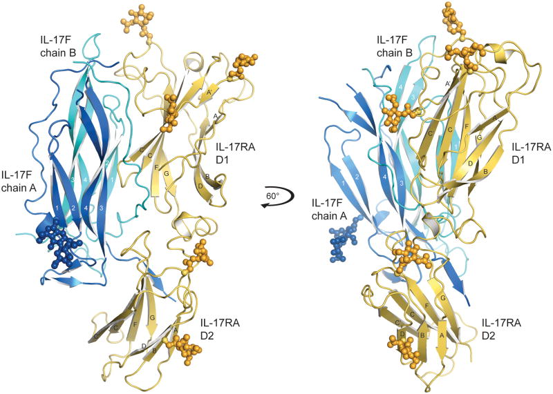
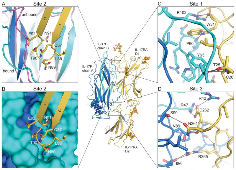
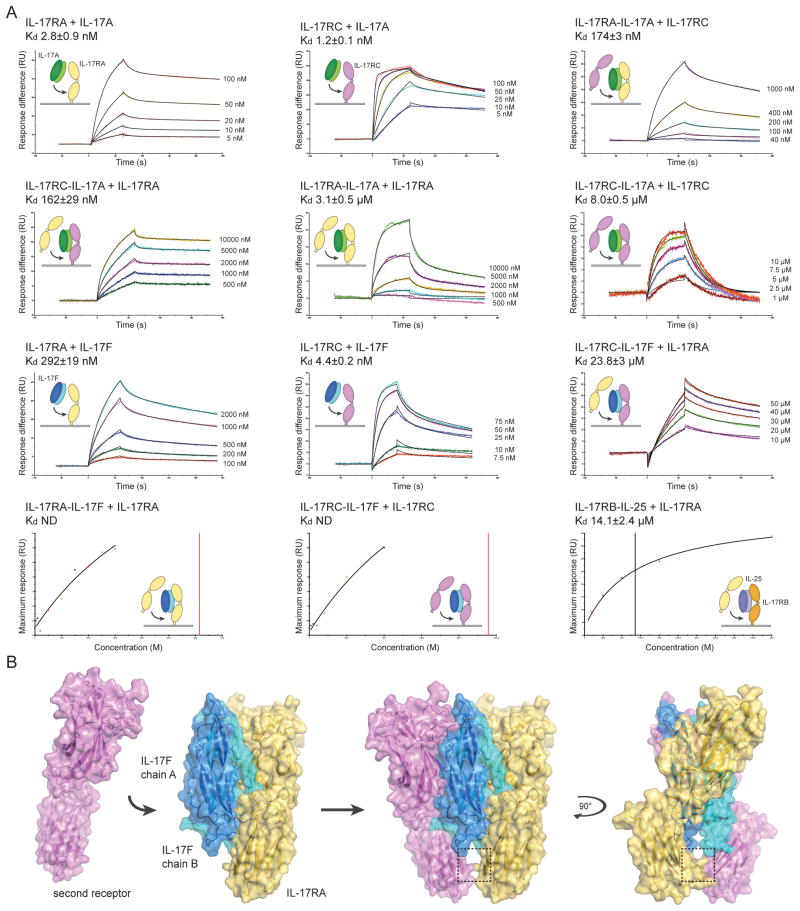
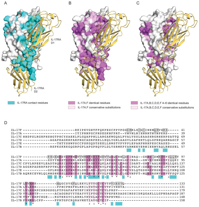
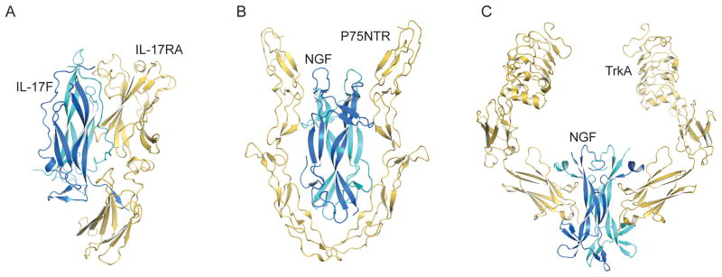
Interleukin 17 (IL-17)-producing helper T cells (T(H)-17 cells), together with their effector cytokines, including members of the IL-17 family, are emerging as key mediators of chronic inflammatory and autoimmune disorders. Here we present the crystal structure of a complex of IL-17 receptor A (IL-17RA) bound to IL-17F in a 1:2 stoichiometry. The mechanism of complex formation was unique for cytokines and involved the engagement of IL-17 by two fibronectin-type domains of IL-17RA in a groove between the IL-17 homodimer interface. Binding of the first receptor to the IL-17 cytokines modulated the affinity and specificity of the second receptor-binding event, thereby promoting heterodimeric versus homodimeric complex formation. IL-17RA used a common recognition strategy to bind to several members of the IL-17 family, which allows it to potentially act as a shared receptor in multiple different signaling complexes.


Conflict of interest statement

Conflict of interest

K.C.G. declares a competing financial interest, and plans to file a patent to use information gained from this crystal structure to design IL-17 antagonists.

Figures

Figure 1
Figure 1
Structure of the IL-17RA-IL-17F complex. Ribbon diagram of IL-17RA in yellow bound to IL-17F (chain A, blue; chain B, cyan), N-linked glycans are shown in ball-and-stick representation. IL-17RA is composed of two fibronectin type III domains (D1 and D2) joined by a short helical linker. The right-hand panel shows the complex rotated by 60° around the y-axis.
Figure 2
Figure 2
IL-17F binding to IL-17RA is mediated by three distinct interfaces. (A) Site 2, the IL-17RA D1 C–C’ loop (yellow) inserts between the N-terminal coil region and strands 1 and 2 of the IL-17F chain B (cyan). The N-terminal coil undergoes a conformational change between the unbound (magenta) and bound (cyan) conformations. (B) Site 2, surface representation of the knob-in-holes IL-17F binding pocket complementarity. (C) Site 1, the IL-17RA D1 N-terminal binding site. (D) Site 3, the IL-17RA D2 binding site. Contact residues are shown as stick models. Gray dotted lines represent hydrogen bonds and pink dotted lines represent salt-bridges.
Figure 3
Figure 3
Assembly and model of the heterodimeric IL-17 signaling complex. (A) IL-17 receptor-cytokine affinity was measured by surface plasmon resonance (SPR). IL-17RA, IL-17RB and IL-17RC were immobilized on the SPR chip surface, and the binding affinity of IL-17A, IL-17F or IL-25 was measured. Where indicated, the affinity of a second receptor binding to the pre-assembled receptor-cytokine complex on the chip was then measured. For kinetic experiments (top 3 rows), representative SPR sensorgrams are shown as colored lines and the curve-fit as a black line. Time in seconds (s) is plotted against response (RU, resonance units). The injected concentrations are to the right of the sensorgrams. For equilibrium experiments (fourth row), the injected concentration (M) is plotted against the maximum response (RU) for a representative experiment; the curve fit is shown as a black line and the dissociation constant (Kd) is marked as a vertical line. The insets show cartoon representations of the binding event. IL-17RA is colored yellow, IL-17RB in orange, IL-17RC in magenta, IL-17A in dark and light green, IL-17F in dark and light blue and IL-25 in dark and light purple. The Kd is reported as the mean of at least two independent experiments ± the standard error of the mean. (B) Model of heterodimeric signaling complex formation. The second receptor (magenta) was modeled assuming that both receptors bind to IL-17F in the same orientation. The C-terminal domains (D2) of the receptors come into close proximity as highlighted by the box (see Supplementary Fig. 3 for more details).
Figure 4
Figure 4
Binding interface and conserved IL-17 residues. Surface representation of IL-17F in white with IL-17RA in ribbon format colored yellow. (A) IL-17RA-IL-17F contact residues highlighted in cyan. (B) Residues conserved among IL-17A and IL-17F are mapped onto the IL-17F structure; identical residues are colored magenta and conservative substitutions in light pink. (C) Residues identical among 4, 5 or 6 IL-17 cytokine family members are colored magenta and conservative substitutions across all six cytokines in light pink. (D) Alignment of human IL-17 cytokines. Residues that form contacts in the IL-17RA-IL-17F structure are highlighted by a black box on the IL-17F sequence and in cyan underneath the alignment. Residues that are identical in 4, 5 or 6 cytokines are highlighted in magenta; those identical in all 6 cytokines are also marked with ‘*’; conserved groups are colored light pink and marked with ‘:’.
Figure 5
Figure 5
IL-17RA-IL-17F receptor complex compared to homodimeric cysteine-knot growth factor receptor complexes. (A) IL-17RA-IL-17F, (B) P75NTR-NGF and (C) TrkA-NGF are shown as ribbon models with the receptors in yellow and the cytokines and growth factors in blue and cyan.

References

    1. Mosmann TR, Coffman RL. TH1 and TH2 cells: different patterns of lymphokine secretion lead to different functional properties. Annu Rev Immunol. 1989;7:145–173. - PubMed
    1. Harrington LE, et al. Interleukin 17-producing CD4+ effector T cells develop via a lineage distinct from the T helper type 1 and 2 lineages. Nat Immunol. 2005;6:1123–1132. - PubMed
    1. Park H, et al. A distinct lineage of CD4 T cells regulates tissue inflammation by producing interleukin 17. Nat Immunol. 2005;6:1133–1141. - PMC - PubMed
    1. Korn T, Bettelli E, Oukka M, Kuchroo VK. IL-17 and Th17 Cells. Annu Rev Immunol. 2009;27:485–517. - PubMed
    1. Weaver CT, Hatton RD, Mangan PR, Harrington LE. IL-17 family cytokines and the expanding diversity of effector T cell lineages. Annu Rev Immunol. 2007;25:821–852. - PubMed

Publication types

Associated data