. 2009 Nov;15(11):1266-72.
doi: 10.1038/nm.2048. Epub 2009 Oct 18.

PPAR-delta senses and orchestrates clearance of apoptotic cells to promote tolerance

Affiliations

PPAR-delta senses and orchestrates clearance of apoptotic cells to promote tolerance

Lata Mukundan et al. Nat Med. 2009 Nov.

Abstract

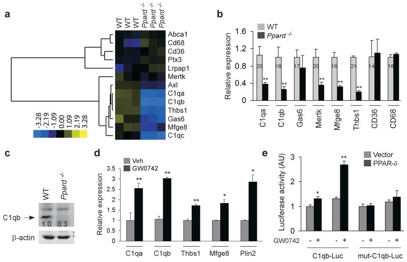
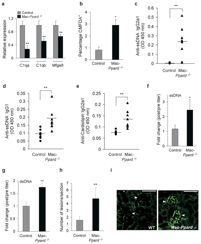
Macrophages rapidly engulf apoptotic cells to limit the release of noxious cellular contents and to restrict autoimmune responses against self antigens. Although factors participating in recognition and engulfment of apoptotic cells have been identified, the transcriptional basis for the sensing and the silent disposal of apoptotic cells is unknown. Here we show that peroxisome proliferator-activated receptor-delta (PPAR-delta) is induced when macrophages engulf apoptotic cells and functions as a transcriptional sensor of dying cells. Genetic deletion of PPAR-delta decreases expression of opsonins such as complement component-1qb (C1qb), resulting in impairment of apoptotic cell clearance and reduction in anti-inflammatory cytokine production. This increases autoantibody production and predisposes global and macrophage-specific Ppard(-/-) mice to autoimmune kidney disease, a phenotype resembling the human disease systemic lupus erythematosus. Thus, PPAR-delta has a pivotal role in orchestrating the timely disposal of apoptotic cells by macrophages, ensuring that tolerance to self is maintained.


Figures

Figure 1
Figure 1
PPAR-δ orchestrates timely disposal of apoptotic cells. (a-c) Apoptotic cell feeding induces PPAR-δ expression in macrophages. Apoptotic thymocytes were fed to wild-type BMDMs (5:1), and expression of PPAR-δ mRNA (a) and protein (b) was quantified 6 and 24 hours later, respectively. (c) Intracellular staining confirmed absence of PPAR-δ expression in thymocytes, and its induction in macrophages after apoptotic cell feeding. Isotype control: gray histogram; PPAR-δ: unshaded histograms. (d) PPARd−/− macrophages are impaired in phagocytosis of apoptotic cells. Kinetics of phagocytosis of apoptotic cells in wild-type and PPARd−/− macrophages. Experiments were repeated five to six independent times, and a representative experiment is shown above. (e, f) Clearance of apoptotic thymocytes in vivo. Labelled apoptotic thymocytes were injected intravenously (e) or intraperitoneally (f), and clearance was monitored 4-6 hours later. Delayed clearance of apoptotic thymocytes by splenic (e) and peritoneal macrophages (f) in PPARd−/−mice (n=4-5). Recovery of fluorescently-labelled thymocytes is quantified in panels (e) and (f). Data is presented as mean ± s.e.m. *P < 0.05, **P < 0.01. ACs: apoptotic cells; AU: arbitrary units; WT: wild-type.
Figure 2
Figure 2
PPAR-δ regulates expression of opsonins in macrophages. (a) Clustering analysis of microarray data from WT and PPARd−/− macrophages. Note the dramatic reduction in expression of opsonins in PPARd−/− macrophages. (b) Relative expression of opsonins and receptors in WT and PPARd−/− macrophages, as assessed by qRT-PCR. (c) Reduced expression of C1qb protein in PPARd−/− macrophages. (d) PPAR-δ agonist (GW0742 100nM) induces opsonin gene expression in wild type macrophages (n=3-4). (e) Activation of C1qb promoter by PPAR-δ. C1qb promoter fragments containing or lacking the PPAR response element (C1qb-Luc or mut-C1qb-Luc, respectively) were transfected into CV-1 cells and luciferase activity was assayed 18 hours later. Data is presented as mean ± s.e.m. *P < 0.05, **P < 0.01. Cycle time for the highest expressing gene is indicated inside its corresponding bar.
Figure 3
Figure 3
PPAR-δ regulates phagocytosis of apoptotic cells via secretion of opsonins. (a) Treatment with PPAR-δ ligand enhances phagocytosis of apoptotic cells by splenic macrophages in vivo. Percentage of splenic macrophages (CD11b+) containing CMFDA-fluorescence was quantified 1 hour after intravenous injection of CMFDA-labelled apoptotic thymocytes (n=4-5). (b, c) PPAR-δ regulates pathways of apoptotic cell uptake in human macrophages. Treatment with GW0742 (100 nM) enhances opsonin gene expression (b) and phagocytosis of apoptotic cells (c) in single donor human monocyte-derived macrophages. (d) Decreased levels of C1q in serum of PPARd−/− mice. Circulating C1qb was detected by immunoprecipitating C1q from serum followed by immunoblotting for C1qb. (e) Restoration of phagocytic capacity of PPARd−/− macrophages by serum from wild-type mice. BMDMs were incubated with wild-type or PPARd−/− macrophages for 24 hours prior to assaying their phagocytic capacity for apoptotic cells. (f) Enhancement of apoptotic cell uptake by purified human C1q in PPARd−/− macrophages. Data is presented as mean ± s.e.m. *P < 0.05, **P < 0.01. Cycle time for the highest expressing gene is indicated inside its corresponding bar.
Figure 4
Figure 4
PPAR-δ is a transcriptional sensor of apoptotic cells in macrophages. (a-c) Apoptotic cells enhance their own clearance by activating PPAR-δ in macrophages. Apoptotic cell (AC) feeding induces expression of PPAR-δ target genes in a PPAR-δ-dependent manner (a). Apoptotic cells enhance expression of C1qb protein in wild-type but not PPARd−/− macrophages (b). (c) Apoptotic cell feeding enhances phagocytic capacity of macrophages in a PPAR-δ-dependent manner. (d-f) PPAR-δ is required for the switch from inflammatory to immunosuppressive cytokine secretion when macrophages phagocytose apoptotic cells. Apoptotic cells enhance secretion of IL-10 from LPS-stimulated WT, but not PPARd−/− macrophages (d). Apoptotic cells fail to suppress release of pro-inflammatory cytokines, such as IL-12p40 (e) and TNF-α (f), in PPARd−/− macrophages (compare LPS and LPS/AC samples). Data is presented as mean ± s.e.m. *P < 0.05, **P < 0.01.
Figure 5
Figure 5
Mice lacking PPAR-δ spontaneously develop autoimmune disease. (a-d) Production of autoantibodies. Serum levels of ANA, and antibodies to dsDNA, ssDNA and cardiolipin were measured by ELISAs in 13-15 month old wild-type and PPARd−/− female mice (n=7-9). For ssDNA antibody, ANA and dsDNA antibody, serum from pristane-treated Balb/cJ mice is presented as a positive control for induction of autoimmunity. (e) Increased deposition of immune complexes in glomeruli of PPARd−/− mice. Paraffin-embedded sections were stained with FITC-conjugated antibody to mouse IgG. (f) Increased protein excretion in PPARd−/− mice. Ratio of urinary protein to urinary creatinine was determined in 14 month WT and PPARd−/− female mice (n=5). Data is presented as mean ± s.e.m. *P < 0.05, **P < 0.01 (nonparametric Mann-Whitney U-test). Bar=50 microns.
Figure 6
Figure 6
Impaired clearance of apoptotic cells and increased autoimmunity in Mac-PPARd−/− mice. (a) Reduced expression of opsonins in BMDMs from Mac-PPARd−/− mice. (b) Decreased clearance of labelled apoptotic thymocytes in spleens of Mac-PPARd−/− mice. 60×106 CMFDA-labeled apoptotic thymocytes were injected intravenously in control and Mac-PPARd−/− mice (n=3), and presence of CMFDA-labelled cells was quantified 4 hours later in spleens. (c-e) Increased autoantibody production in young Mac-PPARd−/− female mice. Serum levels of ssDNA antibodies (c, d) and cardiolipin antibodies (e) were quantified by ELISAs in 3-6 month old control and Mac-PPARd−/− mice (n=8/genotype). (f-i) Macrophage-specific PPAR-δ is required for tolerogenic responses to intravenously injected apoptotic cells. Control and Mac-PPARd−/− mice were injected with 10 syngenic apoptotic thymocytes for 4 weeks, and changes in titers of ssDNA (f) and dsDNA (g) autoantibodies were quantified. Histologic and immunofluorescence analysis of kidneys performed 9 months after apoptotic cell injections (12 month old mice) revealed increased perivascular inflammation (h) and immune complex deposition in the glomeruli (i). Data is presented as mean ± s.e.m. *P < 0.05, **P < 0.01.

Comment in

References

    1. Savill J, Fadok V. Corpse clearance defines the meaning of cell death. Nature. 2000;407:784–8. - PubMed
    1. Henson PM, Bratton DL, Fadok VA. The phosphatidylserine receptor: a crucial molecular switch? Nat Rev Mol Cell Biol. 2001;2:627–33. - PubMed
    1. Savill J, Dransfield I, Gregory C, Haslett C. A blast from the past: clearance of apoptotic cells regulates immune responses. Nat Rev Immunol. 2002;2:965–75. - PubMed
    1. Lauber K, Blumenthal SG, Waibel M, Wesselborg S. Clearance of apoptotic cells: getting rid of the corpses. Mol Cell. 2004;14:277–87. - PubMed
    1. Erwig LP, Henson PM. Clearance of apoptotic cells by phagocytes. Cell Death Differ. 2008;15:243–50. - PubMed

Publication types

MeSH terms

Associated data