. 2010 Jan 14;29(2):201-13.
doi: 10.1038/onc.2009.323. Epub 2009 Oct 19.

Notch-1 activates estrogen receptor-alpha-dependent transcription via IKKalpha in breast cancer cells

Affiliations

Notch-1 activates estrogen receptor-alpha-dependent transcription via IKKalpha in breast cancer cells

L Hao et al. Oncogene. .

Abstract

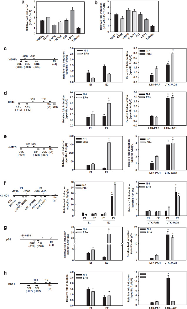
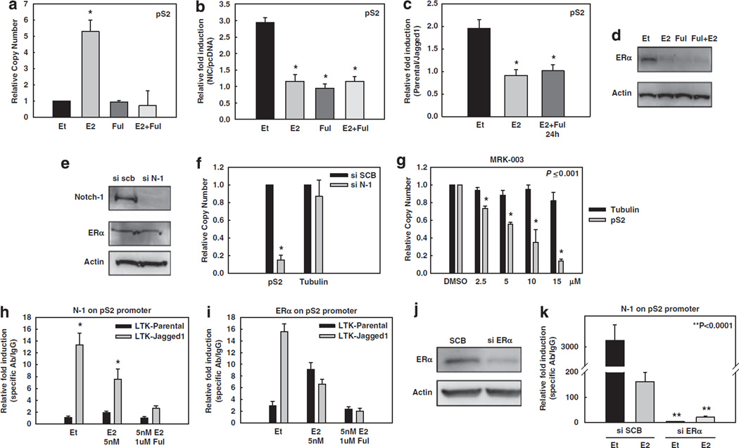
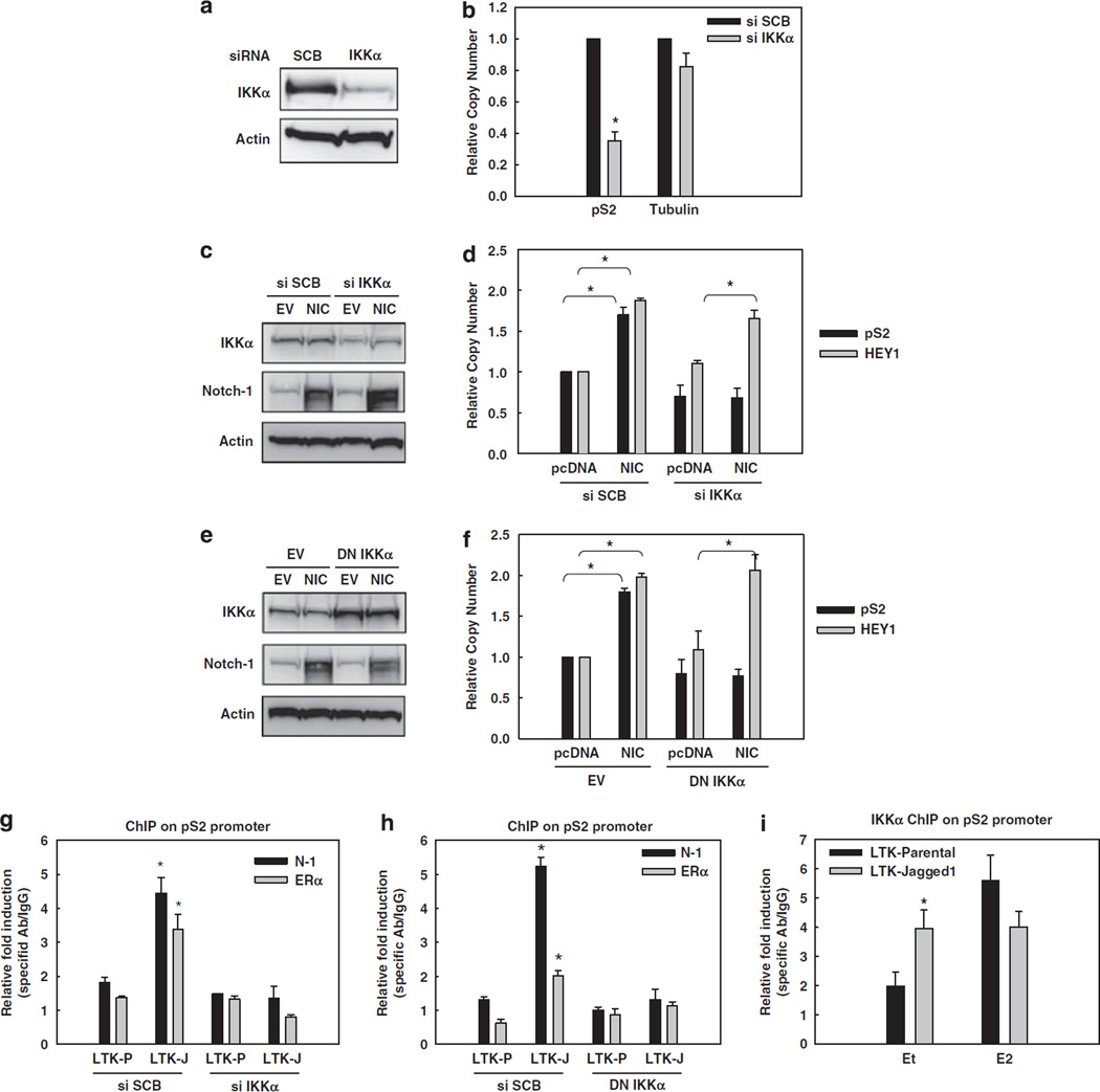
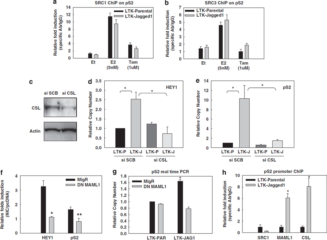
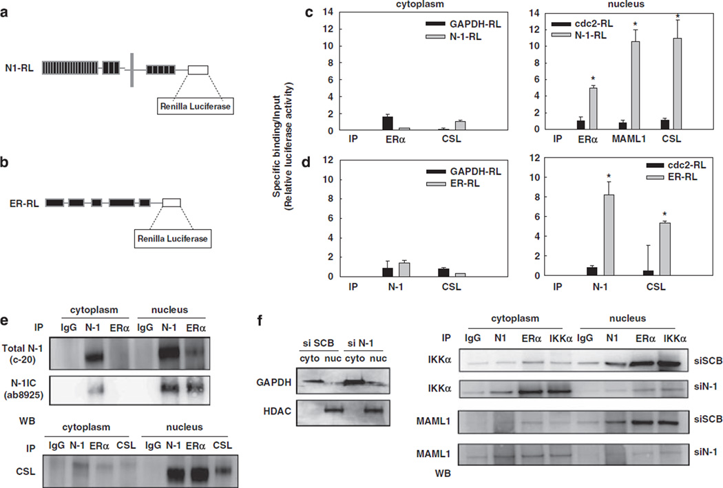
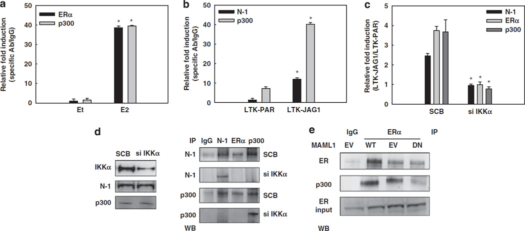
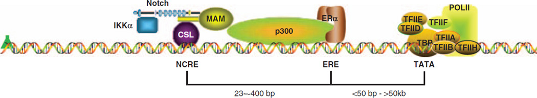
Approximately 80% of breast cancers express the estrogen receptor-alpha (ERalpha) and are treated with anti-estrogens. Resistance to these agents is a major cause of mortality. We have shown that estrogen inhibits Notch, whereas anti-estrogens or estrogen withdrawal activate Notch signaling. Combined inhibition of Notch and estrogen signaling has synergistic effects in ERalpha-positive breast cancer models. However, the mechanisms whereby Notch-1 promotes the growth of ERalpha-positive breast cancer cells are unknown. Here, we demonstrate that Notch-1 increases the transcription of ERalpha-responsive genes in the presence or absence of estrogen via a novel chromatin crosstalk mechanism. Our data support a model in which Notch-1 can activate the transcription of ERalpha-target genes via IKKalpha-dependent cooperative chromatin recruitment of Notch-CSL-MAML1 transcriptional complexes (NTC) and ERalpha, which promotes the recruitment of p300. CSL binding elements frequently occur in close proximity to estrogen-responsive elements (EREs) in the human and mouse genomes. Our observations suggest that a hitherto unknown Notch-1/ERalpha chromatin crosstalk mediates Notch signaling effects in ERalpha-positive breast cancer cells and contributes to regulate the transcriptional functions of ERalpha itself.


Conflict of interest statement

Conflicts of interest

The authors declare no conflict of interest.

Figures

Figure 1
Figure 1
Active Notch-1 facilitates the transcription of ERα-target promoters in the absence of E2. In all experiments, MCF-7 cells were grown in phenol red-free RPMI containing 10% DCC-fetal bovine serum for 3 days prior to harvest. (a) MCF-7 cells were transiently transfected with the active form of Notch-1 (NIC) or pcDNA vector control. The mRNA levels of VEGFα, CD44, c-MYC, CCND1, pS2, HEY1 and β-tubulin were measured by real-time RT–PCR after 48 h after transfection. Values are expressed as relative fold induction by NIC over pcDNA, after internal normalization for 18S rRNA. (b) MCF-7 cells were co-cultured with mouse fibroblasts expressing Jagged-1 (LTK–JAG1) or vector-transfected controls (LTK–PAR) for 12 h prior to harvest. The mRNA levels of VEGFα, CD44, c-MYC, CCND1, pS2, HEY1 and β-tubulin were measured by real-time RT–PCR using validated human-specific primers. Values are expressed as relative fold induction by LTK–JAG1 over LTK–PAR, after internal normalization for RPL13a mRNA. (c–h) The schematics of the indicated promoters and ChIP assays. Charcoal-stripped MCF-7 cells were treated with 5 nm E2 or ethanol (vehicle) for 1 h (left), or co-cultured with LTK cells for 3 h (right) before formaldehyde fixation. ChIP assays were performed with antibodies to Notch-1 or ERα, followed by real-time PCR analysis of the indicated regions of each promoter (arrows). Values are expressed as relative fold increase of specific antibody pull-down over IgG control, after normalization for internal control RPL13a. tts, transcription start site; *P < 0.001. ChIP, chromatin immunoprecipitation; ERα, estrogen receptor-α; LTK–JAG1, mouse LTK fibroblasts expressing Notched ligand Jagged-1; LTK–PAR, control vector transduced parental fibroblast; NIC, active form of Notch-1 (Notch-1IC); RPMI, Rosewell Park Memorial Institute; RT–PCR, reverse transcription–PCR; VEGF, vascular endothelial growth factor.
Figure 2
Figure 2
Notch activation increases ER-dependent transcription. In all experiments, MCF-7 cells were grown in phenol red-free RPMI containing 10% DCC-fetal bovine serum for 3 days prior to harvest. (a–g) pS2 real-time RT–PCR experiments: (a) Untransfected MCF-7 cells were treated with 5 nm of E2 (4 h), 1 µm fulvestrant (24 h) or the combination before harvest. Data are expressed as relative copy number normalized to internal control (18S rRNA); *P ⩽ 0.001. (b) Twelve hours after serum starvation, MCF-7 cells were transfected with NIC or pcDNA vector control. Cells were treated with 5 nm of E2 (4 h), 1 µM fulvestrant (24 h) or a combination of both before harvest. Data are expressed as relative fold induction of NIC over pcDNA after normalization to the internal control 18S rRNA; *P ⩽ 0.001. (c) MCF-7 cells were treated with E2 alone or in combination with fulvestrant as described above, and co-cultured with LTK–JAG1 cells for 12 h. Data are expressed as relative fold induction by LTK–JAG1 over LTK–PAR cells after normalization to internal control RPL13a; *P ⩽ 0.001. (d) Western blot of ERα after the treatments described above. (e) Western blot of MCF-7 cells transfected with Notch-1 siRNA or scrambled control (SCB). (f) MCF-7 cells transfected with Notch-1 siRNA or SCB; *P ⩽ 0.001. (g) MCF-7 cells were treated with increasing concentrations of GSI for 24 h. Data are expressed as relative copy number normalized to internal control (18S rRNA). (h, i) ChIP assay on the pS2 promoter. MCF-7 cells were treated with 5 nm E2, ethanol control for 1 h or E2 in combination with 1 µm fulvestrant (24 h) after 3 days of charcoal stripping and co-cultured with LTK–JAG1 cells for 3 h. Data expressed as relative fold increase of specific antibody over IgG control, after normalization to internal control RPL13a. (j) MCF-7 cells were grown in charcoal-stripped media for 3 days and transfected with ERα siRNA or SCB. The western blot shows efficient downregulation of ERα. Actin was used as a loading control. (k) ChIP assay on the pS2 promoter with cells transfected with siRNA to ERα (as described above), co-cultured with LTK–JAG fibroblasts for 3 h and treated with 5 nm E2 or ethanol for 1 h. ChIP, chromatin immunoprecipitation; ERα, estrogen receptor-α; GSI, γ-secretase inhibitor; LTK–JAG1, mouse LTK fibroblasts expressing Notched ligand Jagged-1; LTK–PAR, control vector-transduced parental fibroblast; NIC, active form of Notch-1 (Notch-1IC); RPMI, Rosewell Park Memorial Institute; RT–PCR, reverse transcription–PCR; siRNA, short interfering RNA.
Figure 3
Figure 3
Notch-1 requires IKKα and its kinase activity for the transcriptional activation of ER-dependent genes. In all experiments, MCF-7 cells were grown in charcoal-stripped medium for a total of 3 days. (a) MCF-7 cells were transfected with IKKα siRNA. The expression level of IKKα was measured by western blotting. (b) pS2 real-time RT–PCR was performed with the same cells as in panel a. Data are expressed as relative copy number after normalization to internal control 18 S rRNA. Tubulin was used as negative control; *P < 0.0001. (c) MCF-7 cells were co-transfected with IKKα siRNA or scrambled control and the construct expressing NIC or pcDNA control. Overexpression of NIC and downregulation of IKKα were validated by western blotting, using actin as loading control. (d) HEY1 and pS2 real-time RT–PCR were performed with the same cells as in panel c; * P ⩽ 0.001. (e) MCF-7 cells were co-transfected with DN-IKKα (AA) or the empty vector and NIC or pcDNA control. Overexpression of DN-IKKα (AA) and Notch-1 was validated by western blotting, using actin as loading control. (f) HEY1 and pS2 real-time RT–PCR were performed with the same cells as in panel e; *P ⩽ 0.001. (g, h) MCF-7 cells were transfected with IKKα siRNA (g) or DN-IKKα (h) and co-cultured with LTK–JAG1 or LTK–PAR fibroblasts for 3 h. ChIP assays were performed with antibodies to Notch-1 or ERα, followed by real-time PCR analysis of the pS2 promoter; *P ⩽ 0.001. (i) MCF-7 cells were co-cultured with LTK–JAG1 or LTK–PAR fibroblasts for 3 h and treated with 5 nm E2 or ethanol control for 1 h prior to harvest. ChIP assay was performed with IKKα antibody followed by real-time PCR for the pS2 promoter; *P ⩽ 0.001. ChIP, chromatin immunoprecipitation; DN-IKKα, the dominant-negative form of IKKα; ERα, estrogen receptor-α; GSI, γ-secretase inhibitor; LTK–JAG1, mouse LTK fibroblasts expressing Notched ligand Jagged-1; LTK–PAR, control vector-transduced parental fibroblast; NIC, active form of Notch-1 (Notch-1IC); RT–PCR, reverse transcription–PCR.
Figure 4
Figure 4
SRC-1 or SRC-3 are not required, but CSL and MAML1 are indispensable for the effect of Notch-1 on the pS2 promoter. In all experiments, MCF-7 cells were grown in charcoal-stripped medium for a total of 3 days. SRC-1 (a) and SRC-3 (b) were detected by ChIP on the pS2 promoter. Charcoal-stripped MCF-7 cells were co-cultured with LTK–JAG1 or LTK–PAR fibroblasts for 3 h, and treated with 5 nm E2 (1 h) or 1 µm 4-hydroxytamoxifen (24 h); *P ⩽ 0.001. (c) MCF-7 cells were transfected with CSL siRNA or scrambled control. CSL knockdown was verified by western blotting. (d, e) HEY1 and pS2 real-time RT–PCR was performed with cells transfected with CSL siRNA and co-cultured with LTK fibroblasts. (f) MCF-7 cells were co-transfected with NIC or pcDNA control and DN-MAML1 or the empty vector MigR. HEY1 and pS2 real-time RT–PCR was performed 48 h after transfection. Data are expressed as relative fold induction by NIC over pcDNA (*P ⩽ 0.001, **P ⩽ 0.005). (g) MCF-7 cells were transfected with DN-MAML1 or the empty vector MigR under charcoal-stripped conditions and co-cultured with LTK cells for 12 h. pS2 mRNA level was measured by real-time RT–PCR; *P ⩽ 0.001. (h) ChIP–PCR of SRC1, MAML1, CSL on pS2 promoter with MCF-7 cells co-cultured with LTK–JAG1 or LTK–PAR fibroblasts; *P ⩽ 0.001. ChIP, chromatin immunoprecipitation; DN-MAML1, the dominant-negative form of MAML1; GSI, g-secretase inhibitor; LTK–JAG1, mouse LTK fibroblasts expressing Notched ligand Jagged-1; LTK–PAR, control vector-transduced parental fibroblast; NIC, active form of Notch-1 (Notch-1IC); RT–PCR, reverse transcription–PCR; siRNA, short interfering RNA; SRC, steroid receptor coactivator.
Figure 5
Figure 5
The Notch-1 and ERα transcriptional complexes interact in chromatin-enriched nuclear extracts. (a, b) Schematics for the constructs of N1-RL (a) and ERα-RL (b). (c) MCF-7 cells were transfected with N1-RL or the negative controls GAPDH-RL (for cytoplasmic proteins) and Cdc2-RL (for nuclear proteins). Cells were harvested after 3 days E2 starvation and nuclear extraction was performed. Cytoplasmic or nuclear extracts were immunoprecipitated with ERα, CSL and MAML1 antibodies. (d) MCF-7 cells were transfected with ERα-RL or negative controls. Cells were harvested after 3 days E2 starvation and nuclear extraction was performed. Cytoplasm lysates or nuclear extracts were immunoprecipitated with Notch-1 and CSL antibodies; *P ⩽ 0.001. (e) Standard IP-western blot was performed on cytoplasmic or nuclear extracts from MCF-7 cells grown in charcoal-stripped medium for 3 days with antibodies to Notch-1, ERα, and CSL. (f) MCF-7 cells were grown in charcoal-stripped medium for a total of 3 days. Cells were transfected with Notch-1 siRNA or scrambled control, and harvested 48 h after transfection. The efficiency of nuclear extraction was verified by western blotting (left). IP–western blot was performed with antibodies to Notch-1, ERα and IKKα. A 10-µl volume of eluted material from each IP was analysed by western blotting for IKKα or MAML1. ChIP, chromatin immunoprecipitation; ERα, estrogen receptor-α; ERα-RL, Renilla luciferase-tagged ERα; GAPDH, glyceraldehyde-3-phosphate dehydrogenase; N1-RL, Renilla luciferase-tagged Notch-1; siRNA, short interfering RNA.
Figure 6
Figure 6
Notch activation recruits p300 to the pS2 promoter. (a) ChIP on the pS2 promoter from MCF-7 cells treated with 5 nm E2 for 1 h after 3 days charcoal stripping. (b) ChIP on the pS2 promoter from MCF-7 cells co-cultured with LTK–JAG1 or LTK–PAR fibroblasts for 3 h after 3 days charcoal stripping. Data in panels a and b are expressed as relative fold increase of specific antibody over IgG control, after normalization to human-specific internal control RPL13a. (c) ChIP on the pS2 promoter from MCF-7 cells grown in charcoal-stripped medium for a total of 3 days, transfected with IKKα siRNA or scrambled control, and co-cultured with LTK–JAG1 or LTK–PAR fibroblasts for 3 h. Data are expressed as relative fold induction by LTK–JAG1 over LTK–PAR after normalization to IgG control and internal control RPL13a. (d) Right: Co-IP experiments on MCF-7 cells, charcoal-stripped for 3 days and transfected with IKKα siRNA or scrambled control. Left: IKKα knockdown was confirmed by western blotting. (e) Co-IP of ERα and p300 on MCF-7 cells, charcoal-stripped for 3 days and transfected with either wild-type or DN-MAML1, or the empty vector control; *P ⩽ 0.05. ChIP, chromatin immunoprecipitation; DN-MAML1, the dominant-negative form of MAML1; LTK–JAG1, mouse LTK fibro-blasts expressing Notched ligand Jagged-1; LTK–PAR, control vector-transduced parental fibroblast; siRNA, short interfering RNA.
Figure 7
Figure 7
Working model of the Notch–ERα crosstalk. The Notch transcriptional complex (NTC) including Notch-1, CSL, MAML1 (MAM) and other coactivators (not shown for clarity) binds to Notch-CSL-responsive elements (NCRE). IKKα is recruited to the NTC in a Notch-dependent manner, although it is still unclear whether the interaction with Notch is direct and is necessary for formation of the supramolecular complex. ERα is recruited to the nucleus in a Notch-1-dependent manner, binds to its responsive element (ERE) or possibly through other transcription factors or pioneer factors, and is necessary for NTC recruitment to ERα-responsive genes. In the absence of E2, MAML1 recruits p300 to the complex, taking over the functions normally carried out by p160 ERα coactivators. The formation of a supramolecular complex between the NTC and the ERα transcriptional complex contributes to activate transcription of a subset of ERα-responsive genes in the absence of E2, and for some genes also in its presence. The pol-II complex is depicted at the TATA box with its most important components. The angled green arrow indicates transcriptional start. ER, estrogen receptor; ERα, estrogen receptor-α.

Similar articles

Cited by

References

    1. Aguilera C, Hoya-Arias R, Haegeman G, Espinosa L, Bigas A. Recruitment of IkappaBalpha to the hes1 promoter is associated with transcriptional repression. Proc Natl Acad Sci USA. 2004;101:16537–16542. - PMC - PubMed
    1. Albanese C, Wu K, D’Amico M, Jarrett C, Joyce D, Hughes J, et al. IKKalpha regulates mitogenic signaling through transcriptional induction of cyclin D1 via Tcf. Mol Biol Cell. 2003;14:585–599. - PMC - PubMed
    1. Arsura M, Panta GR, Bilyeu JD, Cavin LG, Sovak MA, Oliver AA, et al. Transient activation of NF-kappaB through a TAK1/ IKK kinase pathway by TGF-beta1 inhibits AP-1/SMAD signaling and apoptosis: implications in liver tumor formation. Oncogene. 2003;22:412–425. - PubMed
    1. Artavanis-Tsakonas S, Rand MD, Lake RJ. Notch signaling: cell fate control and signal integration in development. Science. 1999;284:770–776. - PubMed
    1. Baonza A, Freeman M. Control of cell proliferation in the Drosophila eye by Notch signaling. Dev Cell. 2005;8:529–539. - PubMed

Publication types

MeSH terms