

Differential requirement of CAAX-mediated posttranslational processing for Rheb localization and signaling
- PMID: 19838215
- PMCID: PMC2809798
- DOI: 10.1038/onc.2009.336
Differential requirement of CAAX-mediated posttranslational processing for Rheb localization and signaling
Abstract
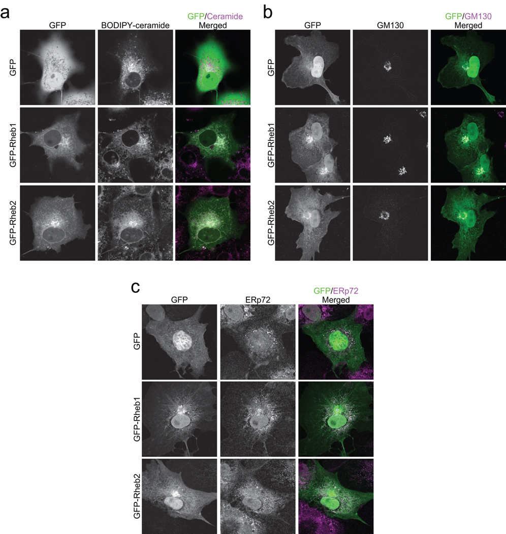
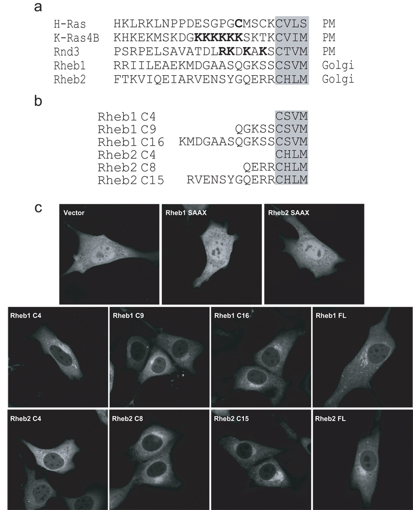
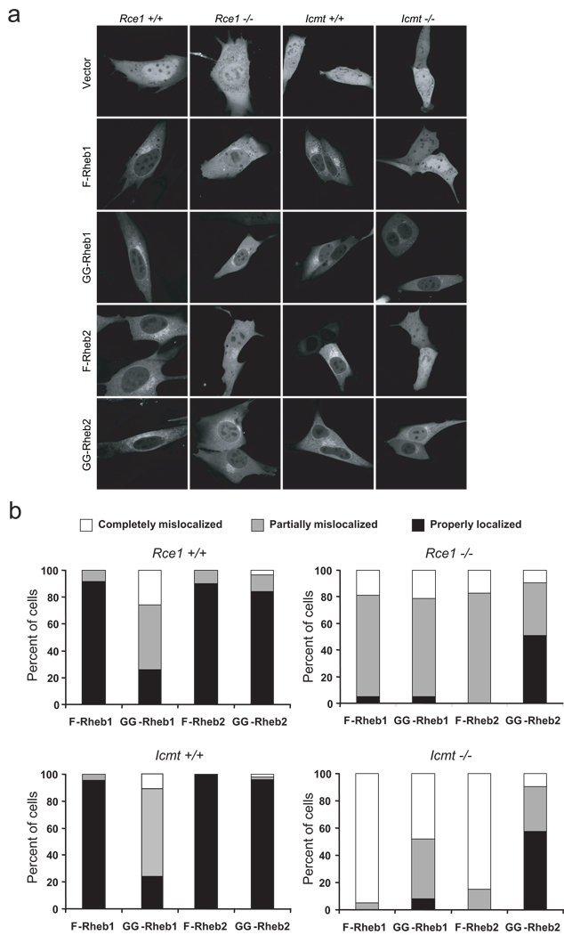
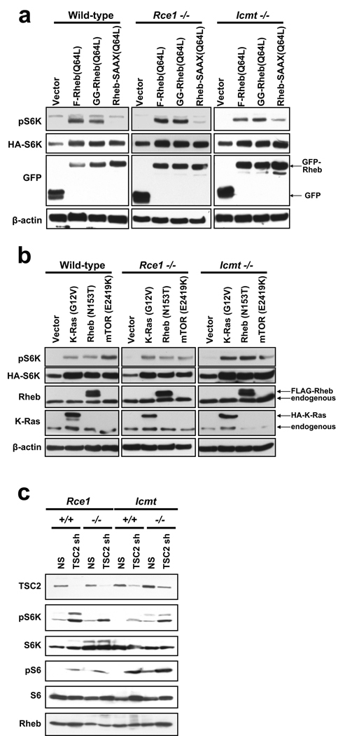
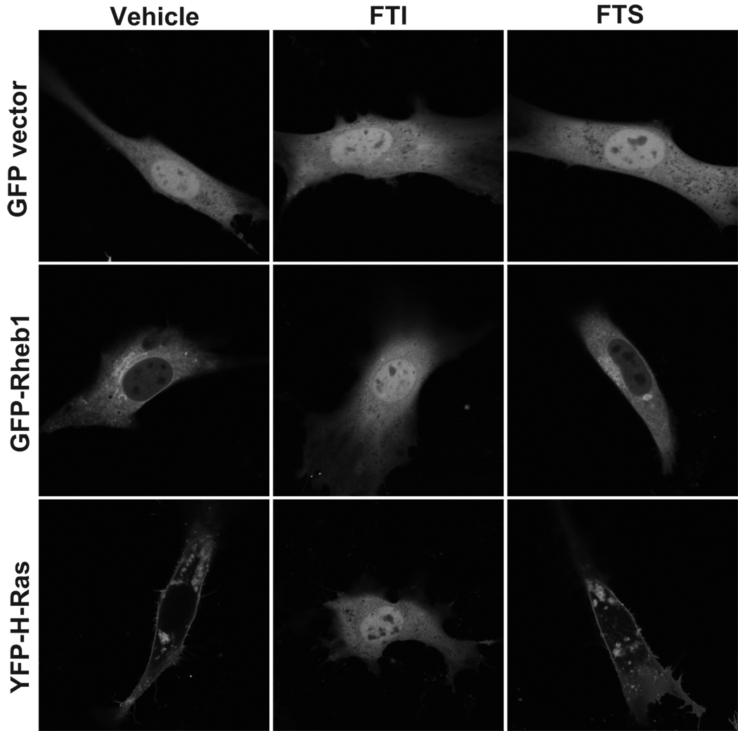
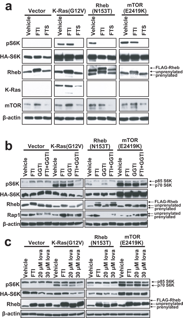
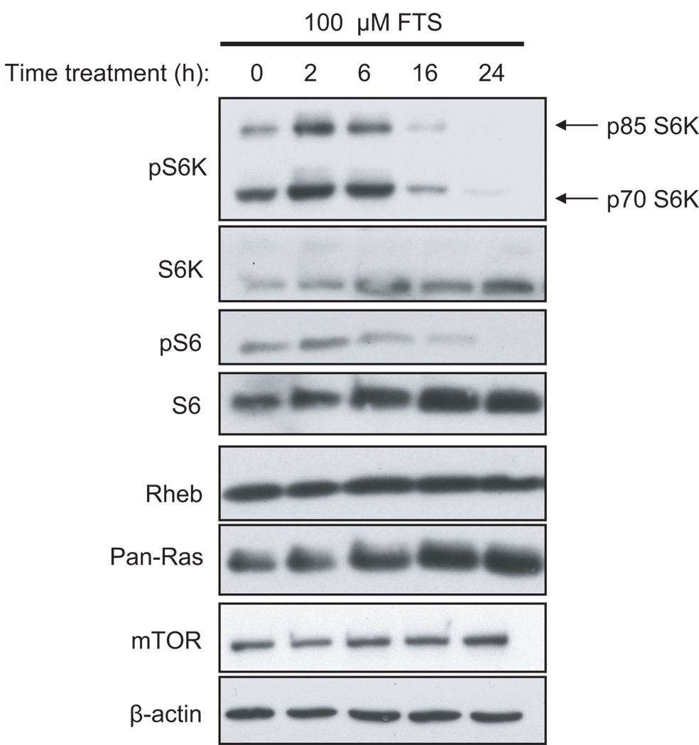
The Rheb1 and Rheb2 small GTPases and their effector mTOR are aberrantly activated in human cancer and are attractive targets for anti-cancer drug discovery. Rheb is targeted to endomembranes via its C-terminal CAAX (C=cysteine, A=aliphatic, X=terminal amino acid) motif, a substrate for posttranslational modification by a farnesyl isoprenoid. After farnesylation, Rheb undergoes two additional CAAX-signaled processing steps, Ras converting enzyme 1 (Rce1)-catalyzed cleavage of the AAX residues and isoprenylcysteine carboxyl methyltransferase (Icmt)-mediated carboxylmethylation of the farnesylated cysteine. However, whether these postprenylation processing steps are required for Rheb signaling through mTOR is not known. We found that Rheb1 and Rheb2 localize primarily to the endoplasmic reticulum and Golgi apparatus. We determined that Icmt and Rce1 processing is required for Rheb localization, but is dispensable for Rheb-induced activation of the mTOR substrate p70 S6 kinase (S6K). Finally, we evaluated whether farnesylthiosalicylic acid (FTS) blocks Rheb localization and function. Surprisingly, FTS prevented S6K activation induced by a constitutively active mTOR mutant, indicating that FTS inhibits mTOR at a level downstream of Rheb. We conclude that inhibitors of Icmt and Rce1 will not block Rheb function, but FTS could be a promising treatment for Rheb- and mTOR-dependent cancers.
Conflict of interest statement
The authors declare no conflict of interest.
Figures
Similar articles
-
Differential membrane localization of ERas and Rheb, two Ras-related proteins involved in the phosphatidylinositol 3-kinase/mTOR pathway.J Biol Chem. 2005 Sep 23;280(38):32768-74. doi: 10.1074/jbc.M506280200. Epub 2005 Jul 26. J Biol Chem. 2005. PMID: 16046393
-
The farnesyl transferase inhibitor (FTI) SCH66336 (lonafarnib) inhibits Rheb farnesylation and mTOR signaling. Role in FTI enhancement of taxane and tamoxifen anti-tumor activity.J Biol Chem. 2005 Sep 2;280(35):31101-8. doi: 10.1074/jbc.M503763200. Epub 2005 Jul 8. J Biol Chem. 2005. PMID: 16006564
-
Farnesylthiosalicylic acid (salirasib) inhibits Rheb in TSC2-null ELT3 cells: a potential treatment for lymphangioleiomyomatosis.Int J Cancer. 2012 Mar 15;130(6):1420-9. doi: 10.1002/ijc.26139. Epub 2011 Aug 1. Int J Cancer. 2012. PMID: 21500191
-
Non-canonical functions of the tuberous sclerosis complex-Rheb signalling axis.EMBO Mol Med. 2011 Apr;3(4):189-200. doi: 10.1002/emmm.201100131. Epub 2011 Mar 16. EMBO Mol Med. 2011. PMID: 21412983 Free PMC article. Review.
-
To cease or to proliferate: new insights into TCTP function from a Drosophila study.Cell Adh Migr. 2007 Jul-Sep;1(3):129-30. doi: 10.4161/cam.1.3.4901. Epub 2007 Jul 16. Cell Adh Migr. 2007. PMID: 19262129 Free PMC article. Review.
Cited by
-
Advances in Autophagy Regulatory Mechanisms.Cells. 2016 May 13;5(2):24. doi: 10.3390/cells5020024. Cells. 2016. PMID: 27187479 Free PMC article. Review.
-
Protein Prenylation and Hsp40 in Thermotolerance of Plasmodium falciparum Malaria Parasites.mBio. 2021 Jun 29;12(3):e0076021. doi: 10.1128/mBio.00760-21. Epub 2021 Jun 29. mBio. 2021. PMID: 34182772 Free PMC article.
-
Direct Interaction between Ras Homolog Enriched in Brain and FK506 Binding Protein 38 in Cashmere Goat Fetal Fibroblast Cells.Asian-Australas J Anim Sci. 2014 Dec;27(12):1671-7. doi: 10.5713/ajas.2014.14145. Asian-Australas J Anim Sci. 2014. PMID: 25358358 Free PMC article.
-
An mTORC1-GRASP55 signaling axis controls unconventional secretion to reshape the extracellular proteome upon stress.Mol Cell. 2021 Aug 19;81(16):3275-3293.e12. doi: 10.1016/j.molcel.2021.06.017. Epub 2021 Jul 9. Mol Cell. 2021. PMID: 34245671 Free PMC article.
-
Bending the path to TOR.Nat Cell Biol. 2010 Nov;12(11):1031-3. doi: 10.1038/ncb1110-1031. Nat Cell Biol. 2010. PMID: 21045803
References
-
- Aspuria PJ, Tamanoi F. The Rheb family of GTP-binding proteins. Cell Signal. 2004;16:1105–1112. - PubMed
-
- Astrinidis A, Henske EP. Tuberous sclerosis complex: linking growth and energy signaling pathways with human disease. Oncogene. 2005;24:7475–7481. - PubMed
-
- Basso AD, Mirza A, Liu G, Long BJ, Bishop WR, Kirschmeier P. The farnesyl transferase inhibitor (FTI) SCH66336 (lonafarnib) inhibits Rheb farnesylation and mTOR signaling. Role in FTI enhancement of taxane and tamoxifen anti-tumor activity. J Biol Chem. 2005;280:31101–31108. - PubMed
-
- Blum R, Cox AD, Kloog Y. Inhibitors of chronically active ras: potential for treatment of human malignancies. Recent Patents Anticancer Drug Discov. 2008;3:31–37. - PubMed
-
- Bodemann BO, White MA. Ral GTPases and cancer: linchpin support of the tumorigenic platform. Nat Rev Cancer. 2008;8:133–140. - PubMed
Publication types
MeSH terms
Substances
Grants and funding
LinkOut - more resources
Full Text Sources
Miscellaneous
