. 2010 May;25(5):1147-56.
doi: 10.1359/jbmr.091031.

Vessel formation is induced prior to the appearance of cartilage in BMP-2-mediated heterotopic ossification

Affiliations

Vessel formation is induced prior to the appearance of cartilage in BMP-2-mediated heterotopic ossification

Christine Fouletier Dilling et al. J Bone Miner Res. 2010 May.

Abstract

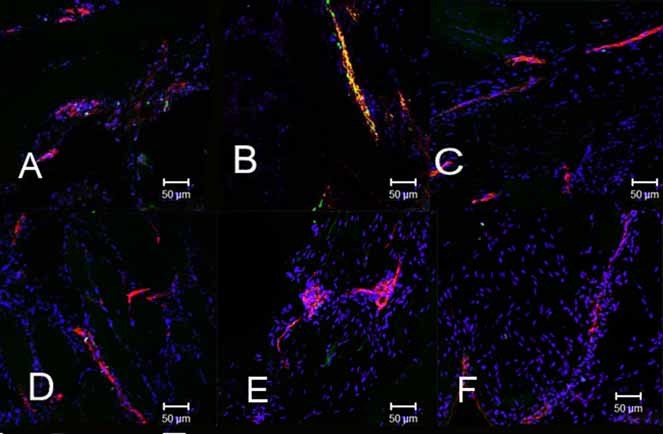
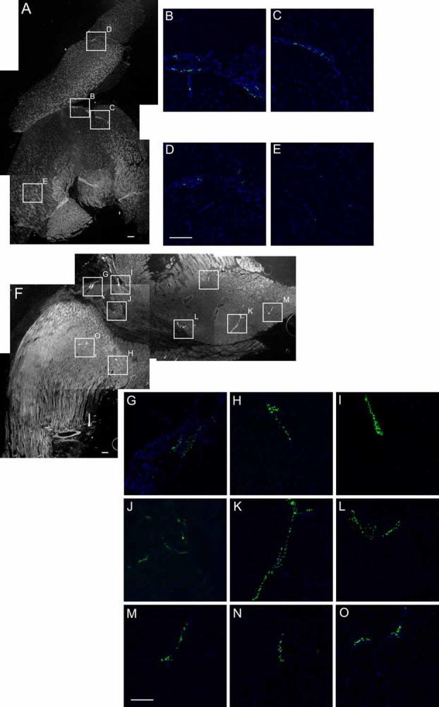
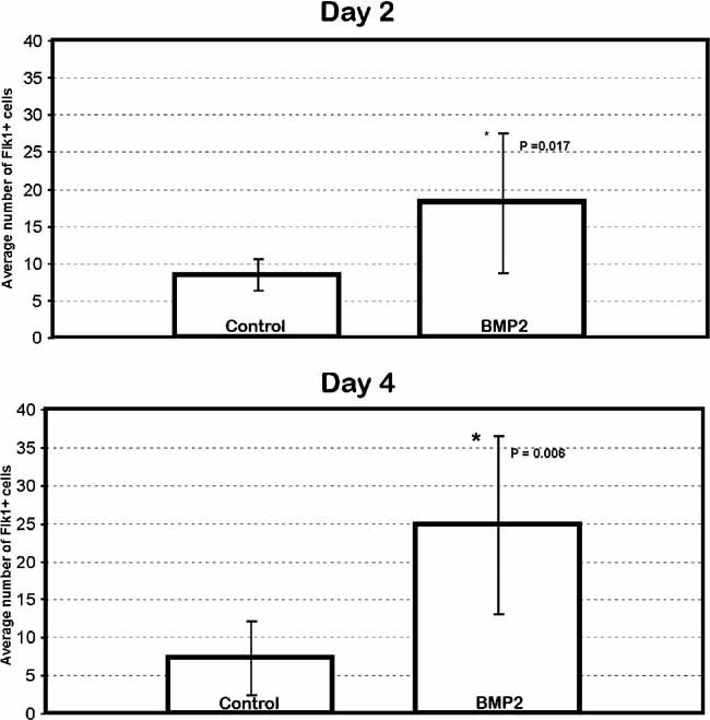
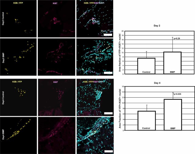
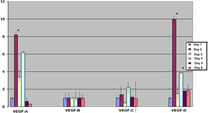
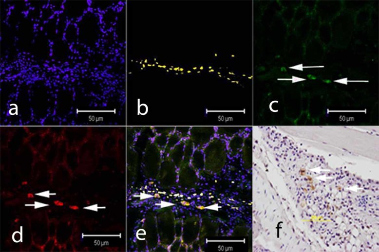
Heterotopic ossification (HO), or endochondral bone formation at nonskeletal sites, often results from traumatic injury and can lead to devastating consequences. Alternatively, the ability to harness this phenomenon would greatly enhance current orthopedic tools for treating segmental bone defects. Thus, understanding the earliest events in this process potentially would allow us to design more targeted therapies to either block or enhance this process. Using a murine model of HO induced by delivery of adenovirus-transduced cells expressing bone morphogenetic protein 2 (BMP-2), we show here that one of the earliest stages in this process is the establishment of new vessels prior to the appearance of cartilage. As early as 48 hours after induction of HO, we observed the appearance of brown adipocytes expressing vascular endothelial growth factors (VEGFs) simultaneous with endothelial progenitor replication. This was determined by using a murine model that possesses the VEGF receptor 2 (Flk1) promoter containing an endothelial cell enhancer driving the expression of nuclear-localized yellow fluorescent protein (YFP). Expression of this marker has been shown previously to correlate with the establishment of new vasculature, and the nuclear localization of YFP expression allowed us to quantify changes in endothelial cell numbers. We found a significant increase in Flk1-H2B::YFP cells in BMP-2-treated animals compared with controls. The increase in endothelial progenitors occurred 3 days prior to the appearance of early cartilage. The data collectively suggest that vascular remodeling and growth may be essential to modify the microenvironment and enable engraftment of the necessary progenitors to form endochondral bone.


Figures

Fig. 1
Fig. 1
Immunohistochemical analysis of endothelial cell replication in tissues isolated at daily intervals after induction of bone formation with cells expressing BMP-2. (A–E) On days 1, 2, 3, 4, and 5, respectively, after injection of BMP-2-producing cells, paraffin sections were prepared and stained with an antibody against Ki67, followed by a secondary antibody conjugated to Alexa fluor 488 (green) mixed with an anti–von Willibrand Factor (vWF) antibody, followed by a secondary antibody conjugated to Alexa fluor 547 (red). (F) A representative image, similar staining, taken from tissues isolated from mice injected with cells transduced with a control vector (Ad5-empty).
Fig. 2
Fig. 2
Wide-field and confocal images of whole tissue sections and quantification of Flk1-H2B::YFP cells. (A, F) Representative montages of low-magnification gray-scale images (1 pixel = 0.003 mm) used for calculating total area for tissue sections. A single representative tissue section is depicted after the entire hind limb muscles that encompassed the injection site were isolated 2 days after receiving an intramuscular injection of cells transduced with either Ad5-empty control vector (A) or Ad5-BMP-2 (B) and sectioned at 15 µm thickness. Although every fifth section across the entire tissue was analyzed, we show only a single representative image of each type. The corresponding regions with positive YFP signal, shown by the boxed areas, were imaged by confocal microscopy (B–E, G–O) for counting the YFP+ cell numbers.
Fig. 3
Fig. 3
Increase in Flk-H2B::YFP+ cells in BMP-2-induced tissue on days 2 and 4. Quantification of Flk1-H2B::YFP+ cells within the tissues 2 and 4 days after induction with Ad5-BMP-2-transduced or control cells. YFP+ nuclei were counted and reported as a ratio of the total area of the tissue section determined using the wide-field montage. Flk-H2B::YFP+ cells were significantly elevated in the tissues receiving BMP-2 compared with controls. The graph depicts the average number of Flk-H2B::YFP+ cells in five sections for day 2 control, 7 sections for day 2 BMP, 8 sections for day 4 control, and 6 sections for day 4 BMP. The number of images taken in each section ranged from 4 to 22. *Denotes a significant difference as determined by the Student's t test.
Fig. 4
Fig. 4
Quantification of YFP+ cell proliferation. Representative images of Flk1-H2B::YFP and the cell proliferation marker Ki67. Colocalization of Flk1-H2B::YFP (yellow) and Ki67 (red) was detected in BMP-2-treated and control tissues. Graphs show the total number of YFP+/Ki67+ cells in the images taken within the area divided by the number of images analyzed. The area fraction was measured for nine at day 2 and five at day 4 BMP and eight at day 2 and four at day 4 control different areas, and the average area fraction was calculated for control and BMP-treated tissues. The area fractions of YFP+/Ki67+ nuclei in the control and the BMP-treated tissues on day 2 were 7.32 ± 3.26 and 10.20 ± 6.95, respectively. The area fractions for day 4 were 6.97 ± 2.32 and 11.26 ± 2.58 in the control and the BMP-treated tissues. Based on the Student's t test, the p value for the day 2 data was .29 and that for the day 4 data was .035. Taken together, the data showed significant YFP+/Ki67+ population increases by day 4 after the BMP treatment, but on day 2 there were no significant differences in dividing YFP cell population between control and BMP-treated tissues.
Fig. 5
Fig. 5
Expression of VEGF-D during the early stages of endochondral bone formation. Results of qRT-PCR analysis of VEGF-A, -B, -C, and -D mRNA levels in tissues surrounding the lesional site that received either the Ad5-BMP-2- or Ad5-empty-transduced cells isolated at daily intervals for up to 7 days after initial injection. Four biologic replicates were run in triplicate, and the averages were normalized against an internal standard (ribosomal RNA). The samples receiving Ad5-BMP-2-transduced cells then were compared with those obtained from the tissues receiving cells transduced with Ad5-empty cassette virus. Therefore, the graph depicts the fold changes in VEGF RNAs in the BMP-2 samples over time compared with control tissues. Error bars depict ± 1 SD unit. *Denotes samples that had a statistically significant (p < .05) difference from all other samples by the ANOVA test.
Fig. 6
Fig. 6
Immunohistochemical staining for brown adipocytes expressing VEGF-D (green, c) in tissues isolated from the Flk1-H2B::YFP mice 4 days after receiving MC3T3 cells transduced with Ad5-BMP-2. Brown adipocytes were identified as cells expressing uncoupling protein 1 (UCP 1; red, d) and yellow (b) represents the Flk-yfp+ endothelial cells within the muscle. The tissues also were stained with VEGF-D antibodies (c) and counterstained with DAPI (blue, a), which stains the nucleus of cells. A merger of these stains (UCP-1, VEGF-D, and YFP) is shown in panel e. In panel f, a paraffin section taken 4 days after injection of BMP-2-producing cells was stained with an antibody against UCP1, and staining was visualized using 3,3'-diaminobenzidine (DAB) as described previously.(8) No staining was observed on a paraffin section taken 4 days after injection of cells transduced with the empty control vector Ad5-HM4 (data not shown).

References

    1. Fraser ST, Hadjantonakis AK, Sahr KE, et al. Using a histone yellow fluorescent protein fusion for tagging and tracking endothelial cells in ES cells and mice. Genesis. 2005;42:162–171. - PMC - PubMed
    1. Shafer J, Davis AR, Gannon FH, et al. Oxygen tension directs chondrogenic differentiation of myelo-monocytic progenitors during endochondral bone formation. Tissue Eng. 2007;13:2011–2019. - PubMed
    1. Colnot C, Lu C, Hu D, Helms JA. Distinguishing the contributions of the perichondrium, cartilage, and vascular endothelium to skeletal development. Dev Biol. 2004;269:55–69. - PubMed
    1. Maes C, Stockmans I, Moermans K, et al. Soluble VEGF isoforms are essential for establishing epiphyseal vascularization and regulating chondrocyte development and survival. J Clin Invest. 2004;113:188–199. - PMC - PubMed
    1. Gerber HP, Vu TH, Ryan AM, Kowalski J, Werb Z, Ferrara N. VEGF couples hypertrophic cartilage remodeling, ossification and angiogenesis during endochondral bone formation. Nat Med. 1999;5:623–628. - PubMed

Publication types

MeSH terms

Substances