Comparative Study
. 2010 Jan;112(1):150-61.
doi: 10.1111/j.1471-4159.2009.06436.x. Epub 2009 Oct 15.

CaMKII associates with CaV1.2 L-type calcium channels via selected beta subunits to enhance regulatory phosphorylation

Affiliations
Comparative Study

CaMKII associates with CaV1.2 L-type calcium channels via selected beta subunits to enhance regulatory phosphorylation

Sunday A Abiria et al. J Neurochem. 2010 Jan.

Abstract

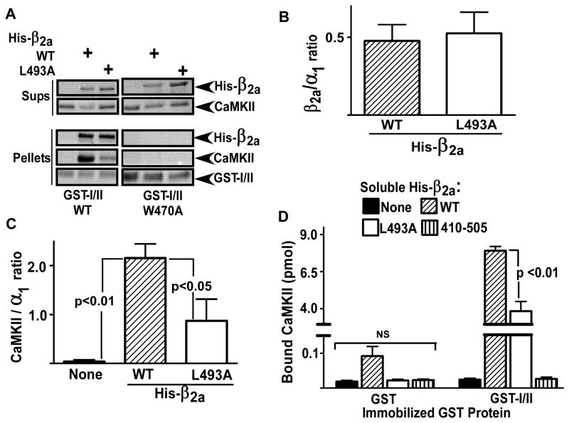
Calcium/calmodulin-dependent kinase II (CaMKII) facilitates L-type calcium channel (LTCC) activity physiologically, but may exacerbate LTCC-dependent pathophysiology. We previously showed that CaMKII forms stable complexes with voltage-gated calcium channel (VGCC) beta(1b) or beta(2a) subunits, but not with the beta(3) or beta(4) subunits (Grueter et al. 2008). CaMKII-dependent facilitation of Ca(V)1.2 LTCCs requires Thr498 phosphorylation in the beta(2a) subunit (Grueter et al. 2006), but the relationship of this modulation to CaMKII interactions with LTCC subunits is unknown. Here we show that CaMKII co-immunoprecipitates with forebrain LTCCs that contain Ca(V)1.2alpha(1) and beta(1) or beta(2) subunits, but is not detected in LTCC complexes containing beta(4) subunits. CaMKIIalpha can be specifically tethered to the I/II linker of Ca(V)1.2 alpha(1) subunits in vitro by the beta(1b) or beta(2a) subunits. Efficient targeting of CaMKIIalpha to the full-length Ca(V)1.2alpha(1) subunit in transfected HEK293 cells requires CaMKII binding to the beta(2a) subunit. Moreover, disruption of CaMKII binding substantially reduced phosphorylation of beta(2a) at Thr498 within the LTCC complex, without altering overall phosphorylation of Ca(V)1.2alpha(1) and beta subunits. These findings demonstrate a biochemical mechanism underlying LTCC facilitation by CaMKII.


Figures

Figure 1
Figure 1. CaMKII associates with LTCC subunits in brain
Triton-soluble extracts of rat (A) or mouse (B-C) forebrains were immunoprecipitated using antibodies to CaMKII (A), β1 (B) or β4 (C) or control IgG. The immune complexes were western blotted for the indicated calcium channel subunits and CaMKII. These data are representative of 2 (A) or 4 (B/C) experiments.
Figure 2
Figure 2. β subunits selectively anchor CaMKII to the Cav1.2α1 I/II linker in vitro
(A) GST-I/II (100 pmol) immobilized in glutathione-coated wells was incubated with a mixture of [32P-T286]CaMKIIα (100 pmol) and either His-β1, His-β2a or His-β3 (36 pmol) in the presence of EDTA to prevent protein phosphorylation. Amounts of bound [32P-T286]CaMKIIα were quantified by scintillation counting and normalized to binding in the presence of β2a. The graph plots data from 3 experiments (mean±sem) analyzed by ANOVA followed by Newman-Keuls multiple comparisons test. (B) Proteins were eluted from the glutathione-coated wells with SDS and western blotted with anti-GST or anti-His antibodies. The blots are representative of the three experiments, and [32P-T286]CaMKIIα binding in this specific experiment is shown below (mean of duplicates).
Figure 3
Figure 3. Molecular determinants of CaMKII anchoring to the Cav1.2α1 I/II linker
(A). Thr286-autophosphorylated CaMKIIα (100 pmol) was incubated with GST-I/II (WT or W470A) (100 pmol) with and without His-tagged β2a (WT or L493A) (100 pmol). Complexes were isolated on glutathione agarose and the bound (pellets) and unbound (sups) proteins were detected by Ponceau-S staining. (B) Quantification (mean±sem, n=3) of β2a(WT) and β2a(L493A) binding to GST-I/II. (C) Quantification (mean±sem, n=3) of CaMKIIα binding to GST-I/II: data were analyzed by one-way ANOVA followed by Newman-Keuls multiple comparisons test. (D) GST or GST-I/II (100 pmol) were immobilized in glutathione-coated wells and incubated with a mixture of [32P-T286]CaMKIIα (100 pmol) and either His-β2a (WT or L493A) or His-β2a(410–505) (100 pmol) in the presence of EDTA. Bound [32P-T286]CaMKIIα was quantified by scintillation counting. Data (mean±sem, n=3) were analyzed by two-way ANOVA with Bonferroni post-test.
Figure 4
Figure 4. The β2a subunit targets CaMKII to Cav1.2α1 subunits in heterologous cells
HEK293 cells co-expressing EGFP-CaMKIIα, HA-Cav1.2α1 and/or FLAG-β2a (WT or L493A) as indicated were incubated for 5 min with A23187 (10 μM). Triton-soluble fractions (Inputs) and anti-HA immune complexes (pellets) were western blotted for HA, FLAG, or EGFP. Unfilled arrowheads indicate a non-specific (NS) FLAG immunoreactive band often detected in HA-immune complexes isolated from cells lacking FLAG-β2a expression that migrates slightly faster than the FLAG-β2a (filled arrowheads). These data are representative of 3 independent experiments.
Figure 5
Figure 5. CaMKII binding does not affect total phosphorylation of Cav1.2α1 and β2a but enhances β2a phosphorylation at Thr498
LTCC complexes were immunoprecipitated from lysates of HEK293 cells co-expressing HA-Cav1.2α1, EGFP-CaMKIIα and FLAG-β2a (WT or L493A) using antibodies to the HA epitope. In panels A and B the cells were labeled with 32P-orthophosphate prior to lysis (see Methods). A. Anti-HA immune complexes were analyzed by SDS-PAGE and autoradiography to detect Cav1.2α1 phosphorylation, and western blotted as indicated. The data are representative of 4 independent experiments. B. Anti-HA immune complexes were analyzed by SDS-PAGE and autoradiography to detect phosphorylation of β2a and were western blotted as indicated. The 32P-phosphate incorporation was quantified and normalized to phosphorylation of wild-type β2a in the absence of EGFP-CaMKIIα (mean±sem, n=4). C. Anti-HA immune complexes were analyzed by western blotting as indicated: blots are representative of 3 similar experiments. The unfilled arrowhead indicates a non-specific (NS) FLAG immunoreactive band detected in HA-immune complexes isolated from cells lacking FLAG-β2a expression that migrates slightly faster than FLAG-β2a (filled arrowhead). D. Quantification of Thr498 phosphorylation of wild-type/mutated FLAG-β2a in anti-HA immune complexes, normalized to phosphorylation of wild-type β2a (mean±sem, n=3). Significance was assessed using a one-sample t-test. CaMKII associates with CaV1.2 L-type calcium channels via selected β subunits to enhance regulatory phosphorylation.

References

    1. Anderson ME, Braun AP, Schulman H, Premack BA. Multifunctional Ca2+/calmodulin-dependent protein kinase mediates Ca(2+)-induced enhancement of the L-type Ca2+ current in rabbit ventricular myocytes. Circ Res. 1994;75:854–861. - PubMed
    1. Berggren PO, Yang SN, Murakami M, et al. Removal of Ca2+ channel beta3 subunit enhances Ca2+ oscillation frequency and insulin exocytosis. Cell. 2004;119:273–284. - PubMed
    1. Bers DM. Calcium cycling and signaling in cardiac myocytes. Annu Rev Physiol. 2008;70:23–49. - PubMed
    1. Birnbaumer L, Qin N, Olcese R, Tareilus E, Platano D, Costantin J, Stefani E. Structures and functions of calcium channel beta subunits. J Bioenerg Biomembr. 1998;30:357–375. - PubMed
    1. Bossuyt J, Helmstadter K, Wu X, Clements-Jewery H, Haworth RS, Avkiran M, Martin JL, Pogwizd SM, Bers DM. Ca2+/calmodulin-dependent protein kinase IIdelta and protein kinase D overexpression reinforce the histone deacetylase 5 redistribution in heart failure. Circ Res. 2008;102:695–702. - PubMed

Publication types

Substances