Review
. 2009 Oct 15;64(1):79-92.
doi: 10.1016/j.neuron.2009.08.038.

Immune influence on adult neural stem cell regulation and function

Affiliations
Review

Immune influence on adult neural stem cell regulation and function

Pamela A Carpentier et al. Neuron. .

Abstract

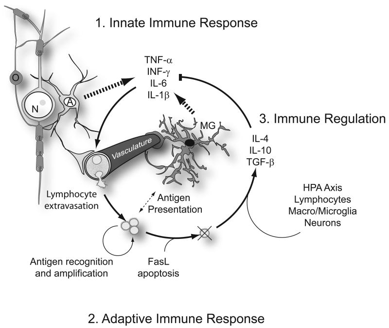
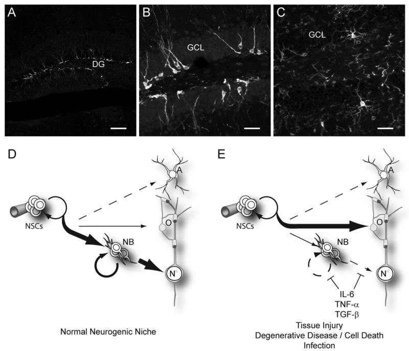
Neural stem cells (NSCs) lie at the heart of central nervous system development and repair, and deficiency or dysregulation of NSCs or their progeny can have significant consequences at any stage of life. Immune signaling is emerging as one of the influential variables that define resident NSC behavior. Perturbations in local immune signaling accompany virtually every injury or disease state, and signaling cascades that mediate immune activation, resolution, or chronic persistence influence resident stem and progenitor cells. Some aspects of immune signaling are beneficial, promoting intrinsic plasticity and cell replacement, while others appear to inhibit the very type of regenerative response that might restore or replace neural networks lost in injury or disease. Here we review known and speculative roles that immune signaling plays in the postnatal and adult brain, focusing on how environments encountered in disease or injury may influence the activity and fate of endogenous or transplanted NSCs.


Figures

Figure 1
Figure 1. Immune response in the CNS
Immune signaling in the CNS follows a well characterized and stereotypical progression. 1. The innate immune response is stimulated as a consequence of infection, injury or cell death, which releases molecules recognized by Toll-like receptors. The initial immune recognition and signaling cascade is mediated primarily by astrocytes (A) and microglia (MG), tissue resident cells that recognize and respond to molecular patterns released by pathogens or damaged cells Astrocytes and microglia are also well known for their intimate contact with the vasculature astrocytic end feet form a tight vascular barrier that modulates immune cell and cytokine trafficking from the periphery. Toll-like receptor activation initiates production of proinflammatory cytokines and chemokines. Key among these are tumor necrosis factor-α (TNF-α), interferon- γ (INF-γ), interleukin-6 (IL-6), and IL-1β. 2. The adaptive immune response is triggered by these cytokines and involves the activation and recruitment of peripheral leucocytes to sites of infection or injury. Cytokines act locally to activate cells of the vasculature as well as within the circulation to activate circulating lymphocytes, which then bind to activated endothelium and extravasate into the site of immune response. Relative to other tissues of the body, peripheral lymphocyte recruitment is attenuated by the astrocytic blood brain barrier (BBB) but injuries that damage the BBB or aggressive acute proinflammatory signaling can degrade BBB function and permit a full-scale adaptive response. Lymphocytes, along with tissue-resident immune cells, then mediate the elimination of pathogens through antigen presentation, recognition and amplification of effector T and B cells. 3. Immune regulation and resolution of the innate and adaptive responses follows clearance of the initiating molecular patterns and Fas-ligand (FasL)-mediated apoptosis of effector lymphocytes. The HPA axis, lymphocytes, macroglia, microglia, and neurons all further contribute to the production of anti-inflammatory hormones and cytokines such as glucocorticoids (via the HPA axis), TGF-β, IL-10, and IL-4.
Figure 2
Figure 2. Impact of proinflammatory signaling on neurogenesis in the adult
A, B. Neurogenesis naturally occurs within the dentate gyrus (DG) of the adult mouse hippocampal formation. Neural stem cells (NSCs) divide to produce progeny that differentiate into an abundant population of doublecortin expressing newborn neurons (white). These immature neurons decorate a thin neurogenic lamina underlying the granular cell layer (GCL). C. As for other regions in the brain, Iba-1-stained microglia (white) in the dentate gyrus send highly ramified processes throughout the tissues, leaving no cell untouched. This intimate association with all cells of the brain allows microglia to constantly and efficiently monitor for infection or cell death and respond by initiating an innate immune response. D. Under normal conditions, the neurogenic niche of the hippocampus strongly favors the production of new neurons (N). This involves first generating a transient amplifying neuroblast (NB) pool, which subsequently differentiate and mature to become integrated within the pre-existing neural networks. Within this region, oligodendrocytes and astrocytes are also produced, though to a much lesser extent. Neurogenesis is supported in this region as well as within the SVZ of the olfactory system by local cues, some of which are provided by cells of the vasculature. In other areas of the brain, oligodendrocytes are the primary product of cycling progenitor cells. E. Upon activation of the innate proinflammatory response, NSC behavior is strongly altered to favor oligodendrocyte production. Of the proinflammatory cytokines released in this initial cascade, IL-6, and TNF-α are well known for their inhibitory effects on neurogenesis. Although TGF-β also plays a role in immune modulation and resolution, it has pleiotropic effects in immune signaling and is also known to impair neurogenesis when chronically over-produced (Buckwalter et al., 2006). In combination, these cytokines impair neuroblasts proliferation as well as reduce survival, maturation, and integration of newly generated postmitotic neurons. (Scale bars = 100 μm in A, 25 μm in B, C)

References

    1. Aarum J, Sandberg K, Haeberlein SL, Persson MA. Migration and differentiation of neural precursor cells can be directed by microglia. Proc Natl Acad Sci U S A. 2003;100:15983–15988. - PMC - PubMed
    1. Agresti C, D'Urso D, Levi G. Reversible inhibitory effects of interferon-gamma and tumour necrosis factor-alpha on oligodendroglial lineage cell proliferation and differentiation in vitro. Eur J Neurosci. 1996;8:1106–1116. - PubMed
    1. Aharonowiz M, Einstein O, Fainstein N, Lassmann H, Reubinoff B, Ben-Hur T. Neuroprotective effect of transplanted human embryonic stem cell-derived neural precursors in an animal model of multiple sclerosis. PLoS ONE. 2008;3:e3145. - PMC - PubMed
    1. Andrews T, Zhang P, Bhat NR. TNFalpha potentiates IFNgamma-induced cell death in oligodendrocyte progenitors. J Neurosci Res. 1998;54:574–583. - PubMed
    1. Arnett HA, Mason J, Marino M, Suzuki K, Matsushima GK, Ting JP. TNF alpha promotes proliferation of oligodendrocyte progenitors and remyelination. Nat Neurosci. 2001;4:1116–1122. - PubMed

LinkOut - more resources