Review
. 2009 Oct 15;64(1):110-22.
doi: 10.1016/j.neuron.2009.08.039.

Immune activation in brain aging and neurodegeneration: too much or too little?

Affiliations
Review

Immune activation in brain aging and neurodegeneration: too much or too little?

Kurt M Lucin et al. Neuron. .

Abstract

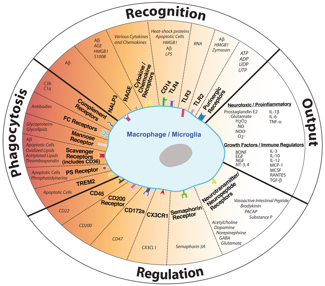
Until recently, the brain was studied almost exclusively by neuroscientists and the immune system by immunologists, fuelling the notion that these systems represented two isolated entities. However, as more data suggest an important role of the immune system in regulating the progression of brain aging and neurodegenerative disease, it has become clear that the crosstalk between these systems can no longer be ignored and a new interdisciplinary approach is necessary. A central question that emerges is whether immune and inflammatory pathways become hyperactivated with age and promote degeneration or whether insufficient immune responses, which fail to cope with age-related stress, may contribute to disease. We try to explore here the consequences of gain versus loss of function with an emphasis on microglia as sensors and effectors of immune function in the brain, and we discuss the potential role of the peripheral environment in neurodegenerative diseases.


Figures

Figure 1
Figure 1. Regulation of microglia/macrophages in the CNS
Microglia/macrophages are activated following the stimulation of various recognition or phagocytic receptors. This activation state is subsequently controlled by neurons, which secrete or express numerous regulatory ligands. The end result of these interactions is the release of cytokines, neurotoxic substrates, and/or growth factors by microglia/macrophages, or the activation of cellular pathways including phagocytosis. Aberrant function of these pathways can result in significant degeneration during aging or disease. For an extensive review of cytokines and chemokines recognized by microglia, refer to (Hanisch, 2002). Aβ, amyloid-β; ADP/ATP, adenosine di/triphosphate; AGE, advanced glycation end product; BDNF, brain-derived neurotrophic factor; EGF, epidermal growth factor; GABA, gamma-aminobutyric acid; H2O2, hydrogen peroxide; HMGB1, High-mobility group box 1; IL-1β, interleukin 1β; LPS, lipopolysaccharide; MCP-1, monocyte chemotactic protein-1; MCSF, macrophage colony-stimulating factor; NGF, nerve growth factor; NO, nitric oxide; NOO, peroxynitrite; NT-3,4, neurotrophin-3,4; O2, superoxide; PACAP, pituitary adenylate cyclase-activating peptide; PS, phosphatidylserine; RAGE, receptor for advanced glycation end products; RANTES, regulated upon activation, normal T cell expressed and secreted; TGF-β, transforming growth factor-β; TLR, toll-like receptor; TNFα, tumour necrosis factor-α; TREM2, triggering receptor expressed by myeloid cells-2; UDP/UTP, uridine di/triphosphate. Receptors displayed inside the cell represent intracellular receptors. Different receptor shapes are not meant to represent actual receptor structures.
Figure 2
Figure 2. Increased microglial activation in the aged brain
Immunostaining for the endosomal/lysosomal enzyme CD68, a marker of the microglia/macrophage lineage, shows a prominent increase in the neocortex of 24-month-old C57BL/6 mouse (B) compared with a 6-month-old mouse (A) consistent with increased microglial activation in aged brains. Inserts show individual microglia under higher magnification where hypertrophied cell bodies are evident in aged brains (compare insert A to B). Scale bars represent 50µm under 20 or 40× magnification.

References

    1. Ajami B, Bennett JL, Krieger C, Tetzlaff W, Rossi FM. Local self-renewal can sustain CNS microglia maintenance and function throughout adult life. Nat Neurosci. 2007;10:1538–1543. - PubMed
    1. Akiyama H, Barger S, Barnum S, Bradt B, Bauer J, Cole GM, Cooper NR, Eikelenboom P, Emmerling M, Fiebich BL, et al. Inflammation and Alzheimer’s disease. Neuroinflammation Working Group. Neurobiol. Aging. 2000;21:383–421. - PMC - PubMed
    1. Aldskogius H, Liu L, Svensson M. Glial responses to synaptic damage and plasticity. J. Neurosci. Res. 1999;58:33–41. - PubMed
    1. Amatniek JC, Hauser WA, DelCastillo-Castaneda C, Jacobs DM, Marder K, Bell K, Albert M, Brandt J, Stern Y. Incidence and predictors of seizures in patients with Alzheimer's disease. Epilepsia. 2006;47:867–872. - PubMed
    1. Arancio O, Zhang HP, Chen X, Lin C, Trinchese F, Puzzo D, Liu S, Hegde A, Yan SF, Stern A, et al. RAGE potentiates Abeta-induced perturbation of neuronal function in transgenic mice. Embo J. 2004;23:4096–4105. - PMC - PubMed

Publication types