Review
. 2009 Oct;13(5):438-45.
doi: 10.1016/j.jaapos.2009.08.011.

Image analysis for retinopathy of prematurity diagnosis

Affiliations
Review

Image analysis for retinopathy of prematurity diagnosis

Michael F Chiang et al. J AAPOS. 2009 Oct.

Abstract

Purpose: To review findings from the authors' published studies involving telemedicine and image analysis for retinopathy of prematurity (ROP) diagnosis.

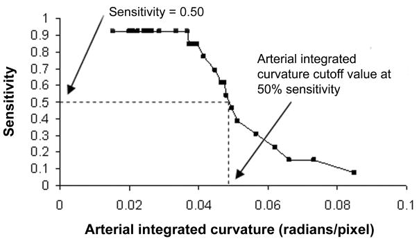
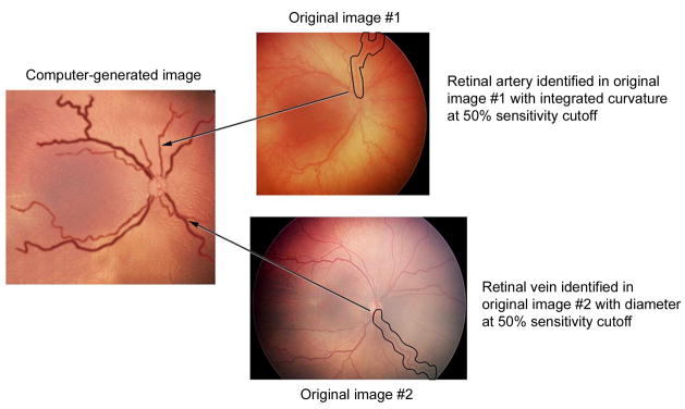
Methods: Twenty-two ROP experts interpreted a set of 34 wide-angle retinal images for presence of plus disease. For each image, a reference standard diagnosis was defined from expert consensus. A computer-based system was used to measure individual and linear combinations of image parameters for arteries and veins: integrated curvature (IC), diameter, and tortuosity index (TI). Sensitivity, specificity, and receiver operating characteristic areas under the curve (AUC) for plus disease diagnosis were determined for each expert. Sensitivity and specificity curves were calculated for the computer-based system by varying the diagnostic cutoffs for arterial IC and venous diameter. Individual vessels from the original 34 images were identified with particular diagnostic cutoffs, and combined into composite wide-angle images using graphics editing software.

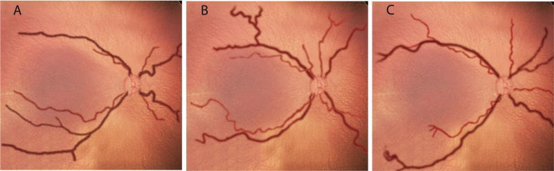
Results: For plus disease diagnosis, expert sensitivity ranged from 0.308-1.000, specificity from 0.571-1.000, and AUC from 0.784 to 1.000. Among computer system parameters, one linear combination had AUC 0.967, which was greater than that of 18 of 22 (81.8%) experts. Composite computer-generated images were produced using the arterial IC and venous diameter values associated with 75% under-diagnosis of plus disease (ie, 25% sensitivity cutoff), 50% under-diagnosis of plus disease (ie, 50% sensitivity cutoff), and 25% under-diagnosis of plus disease (ie, 75% sensitivity cutoff).

Conclusions: Computer-based image analysis has the potential to diagnose severe ROP with comparable or better accuracy than experts, and could provide added value to telemedicine systems. Future quantitative definitions of plus disease might improve diagnostic objectivity.


Figures

FIG 1
FIG 1
Methodology for creating computer-generated images, representing varying levels of vascular abnormality in plus disease. A, Sensitivity cutoff values were determined from study data, in which 34 wide-angle retinal images were reviewed by 22 experts for presence of plus disease. Reference standard diagnosis was defined as consensus of expert responses. “50% sensitivity cutoff” for a vascular parameter (eg, arterial integrated curvature [IC] in this example) is the value associated with 50% under-diagnosis of true plus disease, according to the reference standard. B, Original 34 images were searched to identify vessels (circled) at that sensitivity cutoff for desired parameters (eg, 50% arterial IC and venous diameter). This is repeated for arteries and veins in all quadrants. Vessels are digitally edited into computer-generated image with all vessels at that sensitivity cutoff (eg, 50% in this example).
FIG 1
FIG 1
Methodology for creating computer-generated images, representing varying levels of vascular abnormality in plus disease. A, Sensitivity cutoff values were determined from study data, in which 34 wide-angle retinal images were reviewed by 22 experts for presence of plus disease. Reference standard diagnosis was defined as consensus of expert responses. “50% sensitivity cutoff” for a vascular parameter (eg, arterial integrated curvature [IC] in this example) is the value associated with 50% under-diagnosis of true plus disease, according to the reference standard. B, Original 34 images were searched to identify vessels (circled) at that sensitivity cutoff for desired parameters (eg, 50% arterial IC and venous diameter). This is repeated for arteries and veins in all quadrants. Vessels are digitally edited into computer-generated image with all vessels at that sensitivity cutoff (eg, 50% in this example).
FIG 2
FIG 2
Computer-generated composite images demonstrating arterial integrated curvature and venous diameter values. A, 25% under-diagnosis of true plus disease (ie, 75% sensitivity cutoff). B, 50% under-diagnosis of true plus disease (ie, 50% sensitivity cutoff). C, 75% under-diagnosis of true plus disease (ie, 25% sensitivity cutoff) based on reference standard of consensus among 22 ROP experts. Vessels are superimposed on a choroidal background, with arteries shown in light red and veins in dark red. Vascular appearance was smoothened using graphics-editing software.
FIG 3
FIG 3
Comparison of computer-generated composite images and published example photographs. A, 50% under-diagnosis of true plus disease (ie, 50% sensitivity cutoff). B, standard photographic definition of plus disease selected by expert committee. C, Computer-generated composite image reflecting 25% under-diagnosis of true plus disease (ie, 75% sensitivity cutoff). D, example photograph of pre-plus disease selected by expert committee. Size of composite-generated images adjusted to allow comparison at a similar level of magnification. Reproduced with permission.

References

    1. Palmer EA, Flynn JT, Hardy RJ, et al. Incidence and early course of retinopathy of prematurity: The Cryotherapy for Retinopathy of Prematurity Cooperative Group. Ophthalmology. 1991;98:1628–40. - PubMed
    1. Good WV, Hardy RJ, Dobson V, et al. The incidence and course of retinopathy of prematurity: Findings from the early treatment for retinopathy of prematurity study. Pediatrics. 2005;116:15–23. - PubMed
    1. Martin JA, Hamilton BE, Sutton PD, et al. Births: Final data for 2005. Natl Vital Stat Rep. 2007;56:1–103. - PubMed
    1. National Eye Institute. Facts about retinopathy of prematurity. [Accessed May 6, 2008]. Available at: http://www.nei.nih.gov/health/rop/
    1. Shennan AH, Bewley S. Why should preterm births be rising? BMJ. 2006;332:924–5. - PMC - PubMed

Publication types