. 2009 Dec 18;284(51):35885-95.
doi: 10.1074/jbc.M109.048140.

Aup1-mediated regulation of Rtg3 during mitophagy

Affiliations

Aup1-mediated regulation of Rtg3 during mitophagy

Dikla Journo et al. J Biol Chem. .

Abstract

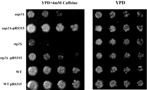
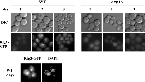
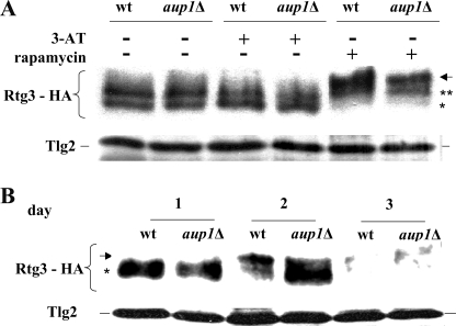
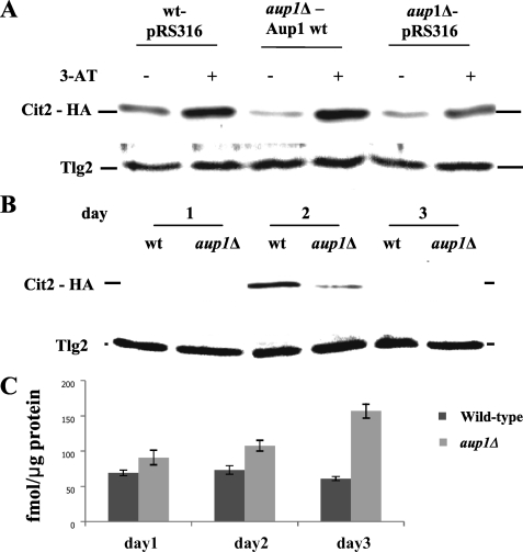
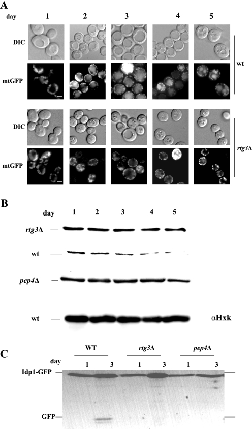
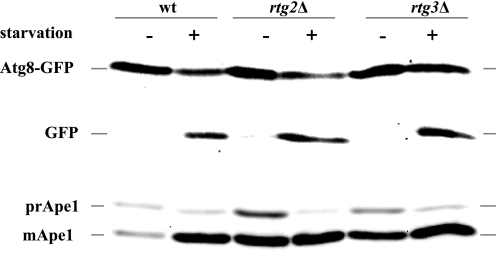
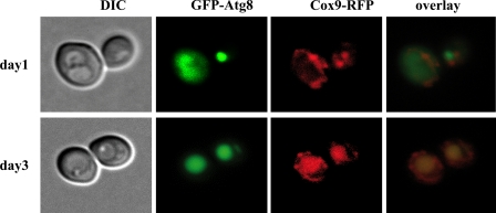
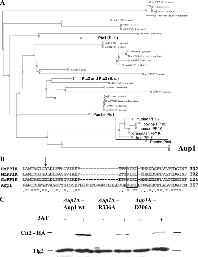
Mitophagy is an autophagic process that degrades mitochondria by an intracellular engulfment that leads to their delivery into the lumen of the cell's hydrolytic compartment, such as the lysosome in animal cells or the vacuole in yeast. It is hypothesized that such processes serve a quality control function to prevent or slow the accumulation of malfunctioning mitochondria, which are thought in turn to underlie central aspects of the aging process in eukaryotic organisms. We recently identified a conserved mitochondrial protein phosphatase homolog, Aup1, which is required for efficient stationary phase mitophagy in yeast. In the present report, we demonstrate that the retrograde signaling pathway (RTG) is defective in aup1Delta mutants. In agreement with a role for Aup1 in the regulation of the RTG pathway, we find that deletion of RTG3, a transcription factor that mediates the RTG response, causes a defect in stationary phase mitophagy and that deletion of AUP1 leads to changes in Rtg3 phosphorylation patterns under these conditions. In addition, we find that mitophagic conditions lead to induction of RTG pathway target genes in an Aup1-dependent fashion. Thus, our results suggest that the function of Aup1 in mitophagy could be explained through its regulation of Rtg3-dependent transcription.


Figures

FIGURE 1.
FIGURE 1.
The caffeine sensitivities of the aup1Δ and rtg2Δ are both suppressed by leucine prototrophy. Wild type (WT) (HAY75), aup1Δ (HAY809), and rtg2Δ (DJY2) were grown to stationary phase in SD medium, serially diluted (from an initial concentration of 6 × 10⁶ cells/ml) in 1:10 steps, and spotted onto YPD + caffeine plates (4 mm). Plates were incubated at 26 °C and imaged. Bottom row of plates, replicate aliquots were serially diluted (from an initial concentration of 6 × 10⁶ cells/ml) in 1:10 steps and spotted onto control YPD plates to demonstrate equivalent growth potential in the absence of caffeine.
FIGURE 2.
FIGURE 2.
Rtg3-GFP is translocated to the nucleus in an Aup1- dependent fashion, during growth on lactate medium. Isogenic wild type (WT) (HAY977) and aup1Δ mutants (HAY984) expressing Rtg3-GFP were grown in SL medium supplemented with 0.2% (w/v) glutamate for 3 days and imaged daily. 4′,6-Diamidino-2-phenylindole (DAPI) staining panels of day 2 wild type cells were added for verification of nuclear localization of the GFP signal. Scale bar, 5 μm. DIC, differential interference contrast.
FIGURE 3.
FIGURE 3.
Aup1 affects Rtg3 phosphorylation during growth on lactate. A, wild type (wt) (DJY8) and aup1Δ (DJY9) cells expressing Rtg3-HA were grown in SD medium supplemented with 0.2% glutamate at 26 °C to an A600 of 0.5 and challenged with 50 mm 3-AT or 0.2 μg/ml rapamycin as indicated. Cells were incubated for 30 min, and 20 μg of protein extracted from each sample were processed for Western blotting. B, cells expressing Rtg3-HA (DJY8) were grown in synthetic lactate medium supplemented with 0.2% glutamate. At each time point, 10 A600 units of cells were sampled, and 20 μg of protein from each sample were analyzed by immunoblotting. The arrow denotes the migration distance of the hyperphosphorylated form in each experiment, whereas asterisks denote the faster migrating (hypophosphorylated) species of Rtg3-HA.
FIGURE 4.
FIGURE 4.
A, Aup1 is required for efficient up-regulation of Cit2 in response to 3-AT and during mitophagy. Wild type (wt) and aup1Δ cells expressing Cit2-HA (DJY3 and DJY4, top) and harboring either empty vector or a vector expressing wild type Aup1 were grown overnight in SD medium lacking histidine and supplemented with 0.2% glutamate to an A600 of 0.5. The cells were then challenged with 50 mm 3-AT for 30 min, and 10 A600 units of cells were sampled at each time point and processed for immunoblotting. 20 μg of protein were loaded per lane, and the filters were probed with anti-HA antibodies to detect Cit2 and with anti-Tlg2 antibodies as load control. B, the RTG pathway was induced during stationary phase mitophagy in an Aup1-dependent fashion. Wild type (DJY3) and aup1Δ (DJY4) cells expressing Cit2-HA were grown in lactate as described in the legend to Fig. 3B, and samples (20 μg of protein) were analyzed by SDS-PAGE and immunoblotting with anti-HA antibody at each time point. Anti-Tlg2 immunoblotting was used as load control. C, oxidative damage accumulates in aup1Δ cells between the second and third days of the incubation, relative to wild type cells. Wild type (HAY75) and aup1Δ (HAY809) cells were grown on lactate medium and sampled as in B. Protein extracts were prepared, and 15 μg of each sample were conjugated to DNPH as described under “Experimental Procedures.” Anti-DNP antibody signals (fmol of DNP incorporated/μg of total protein) were quantified by SDS-PAGE followed by Western blotting. Bars, S.E. (n = 3).
FIGURE 5.
FIGURE 5.
Rtg3 is essential for stationary phase mitophagy. A, AUP1 (HAY805) and aup1Δ (DJY6) cells expressing mitochondrial matrix-targeted GFP (mtGFP) were incubated in synthetic lactate medium for 5 days. Cells were analyzed by fluorescence microscopy daily, and mitophagy was scored as appearance of the GFP signal in the vacuolar lumen. Scale bar, 5 μm. B, wild type (wt) (HAY75), aup1Δ (DJY10), and pep4Δ (TVY1) cells were incubated as in A, and 10 A600 units were sampled daily for anti-aconitase immunoblotting. 20 μg of protein were analyzed per lane. Anti-hexokinase immunoblotting is shown as load control. C, wild type (HAY75), aup1Δ (DJY1), and pep4Δ (TVY1) cells expressing Idp1-GFP were grown as above, and 20 μg of protein were analyzed by immunoblotting with anti-GFP antibody at each time point. Scale bar, 5 μm. DIC, differential interference contrast.
FIGURE 6.
FIGURE 6.
Rtg2 and Rtg3 are not required for starvation-induced autophagy or for the Cvt pathway. Wild type (wt) (HAY75), rtg2Δ (DJY2), and rtg3Δ (DJY5) cells expressing GFP-Atg8 from the TPI promoter (plasmid pYX-GFPATG8) were grown to midlog phase in SD medium, washed, and resuspended in SD-N or in SD for 2 h. Samples (10 A600 units) were then taken and analyzed by immunoblotting with anti-GFP and anti-Ape1 antibodies.
FIGURE 7.
FIGURE 7.
The regulation of mitophagy and starvation-induced macroautophagy can be uncoupled. Wild type cells co-expressing GFP-Atg8 and Cox9-RFP (strain DJY7) were grown in synthetic lactate medium for 3 days. Cells were imaged daily by fluorescence microscopy. DIC, differential interference contrast.
FIGURE 8.
FIGURE 8.
An RXKL motif, conserved between Aup1 and mammalian PP1K proteins, is essential for Aup1 function. A, comparative evolutionary tree for BLAST comparison of Aup1 (in bold, at the bottom; Swissprot designation PP2C-6) against the Swissprot data base. Framed in black are the vertebrate PP1K homologs. Three S. cerevisiae proteins that appear in the comparison are also in bold: Ptc1, Ptc2, and Ptc3. The single non-vertebrate PP2C homolog that shows a higher similarity to Aup1 is Schizosaccharomyces pombe Ptc4, which is an ortholog of the PP1K/Aup1 family. Expansion of the search to the non-redundant data base (which includes unannotated sequences) results in a major expansion of the fungal ortholog clade represented by the single S. pombe Ptc4 node in this picture and a complete loss of S. cerevisiae representatives, suggesting that there are no paralogs of Aup1 within the S. cerevisiae genome and that the PP1K/Aup1 family is a distinct branch within the PP2C protein superfamily. B, identification of a conserved RXKL motif that defines the PP1K family of phosphatase homologs. Human (Hs), mouse (Mm), and dog (Cm) PP1K virtual translation products were aligned with the Aup1 protein sequence using ClustalW. The arrow denotes aspartate 306 in Aup1, which is predicted by threading analysis to participate in the coordination of a catalytically important manganese ion. The RXKL motif is denoted by the black frame. C, aspartate 306 and arginine 336 are essential for Aup1 function. Mutant aup1Δ (HAY 809) cells expressing Cit2-HA were transformed with plasmids encoding wild type, R336A, and D306A variants of AUP1 (see “Experimental Procedures”) expressed under control of the endogenous promoter and tested for their ability to induce the Rtg pathway response gene CIT2 in response to 3-AT. Cells were grown overnight in SD medium lacking histidine and supplemented with 0.2% glutamate to an A600 of 0.5. The cells were then challenged with 50 mm 3-AT for 30 min, and 10 A600 units of cells were sampled at each time point and processed for immunoblotting. 20 μg of protein were loaded per lane, and the filters were probed with anti-HA antibodies and with anti-Tlg2 antibodies as load control.
FIGURE 9.
FIGURE 9.
Arginine 336 of Aup1, within the RXKL motif, is required for phosphatase activity. A, the purified C-terminal PP2C motif iteration (amino acids 213–419) shows PP2C-type phosphatase activity. 2 μg of purified Aup1 (amino acids 213–419) or purified GFP control were assayed for phosphatase activity in the presence of Mg2+ or Mn2+, as detailed under “Experimental Procedures.” B, steady state kinetic analysis of purified Aup1 (amino acids 213–419) versus the D306A and R336A variants. Results are representative of three independent determinations.

Similar articles

Cited by

References

    1. Abeliovich H. (2007) Autophagy 3, 275–277 - PubMed
    1. Wallace D. C. (2005) Annu. Rev. Genet. 39, 359–407 - PMC - PubMed
    1. Abeliovich H., Klionsky D. J. (2001) Microbiol. Mol. Biol. Rev. 65, 463–479 - PMC - PubMed
    1. Klionsky D. J. (2005) Curr. Biol. 15, R282–R283 - PubMed
    1. Klionsky D. J., Emr S. D. (2000) Science 290, 1717–1721 - PMC - PubMed

Publication types

MeSH terms

Substances