. 2009 Dec 11;284(50):34849-60.
doi: 10.1074/jbc.M109.068403. Epub 2009 Oct 19.

GRASP65 and GRASP55 sequentially promote the transport of C-terminal valine-bearing cargos to and through the Golgi complex

Affiliations

GRASP65 and GRASP55 sequentially promote the transport of C-terminal valine-bearing cargos to and through the Golgi complex

Giovanni D'Angelo et al. J Biol Chem. .

Abstract

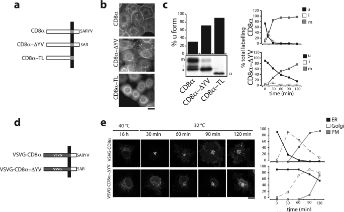
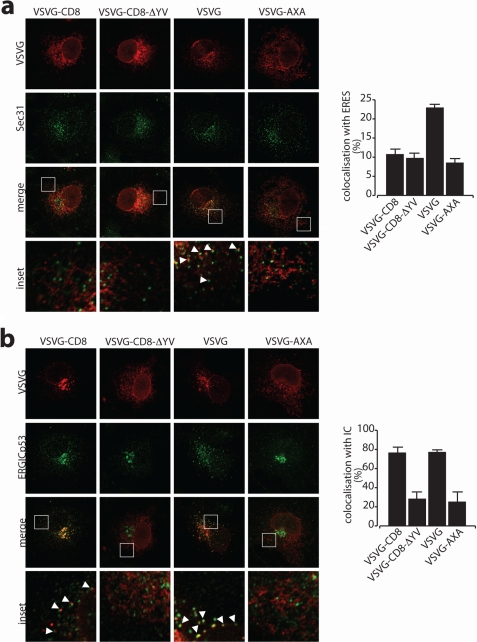
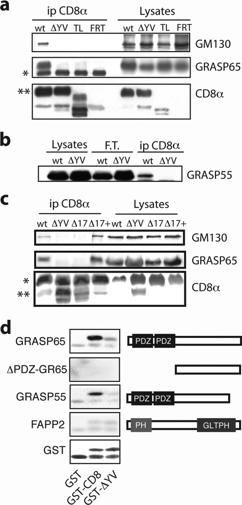
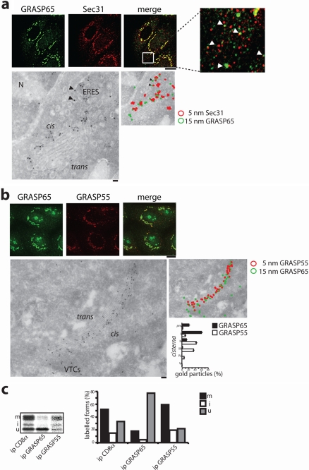
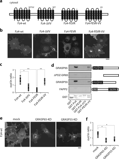
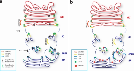
The Golgi matrix proteins GRASP65 and GRASP55 have recognized roles in maintaining the architecture of the Golgi complex, in mitotic progression and in unconventional protein secretion whereas, surprisingly, they have been shown to be dispensable for the transport of commonly used reporter cargo proteins along the secretory pathway. However, it is becoming increasingly clear that many trafficking machineries operate in a cargo-specific manner, thus we have investigated whether GRASPs may control the trafficking of selected classes of cargo. We have taken into consideration the C-terminal valine-bearing receptors CD8alpha and Frizzled4 that we show bind directly to the PSD95-DlgA-zo-1 (PDZ) domains of GRASP65 and GRASP55. We demonstrate that both GRASPs are needed sequentially for the efficient transport to and through the Golgi complex of these receptors, thus highlighting a novel role for the GRASPs in membrane trafficking. Our results open new perspectives for our understanding of the regulation of surface expression of a class of membrane proteins, and suggests the causal mechanisms of a dominant form of autosomal human familial exudative vitreoretinopathy that arises from the Frizzled4 mutation involving its C-terminal valine.


Figures

FIGURE 1.
FIGURE 1.
The C-TVM of CD8α enhances its intracellular transport. a, schematic representations of the three different CD8α constructs used. TL, tail less. b, immunofluorescence analysis of FRT cells stably expressing the CD8α constructs, as indicated. Bar, 10 μm. c, left panels, SDS-PAGE analysis of the immunoprecipitated forms of CD8α and CD8α-ΔYV labeled in stably expressing cells after a 30-min pulse with [35S]Cys/Met. The quantitation of CD8 forms was performed by densitometric scanning and the relative amounts of CD8u (see “Experimental Procedures”) are shown in the histogram. The TL mutant shows a faster mobility on SDS-PAGE due to the deletion of the 28-amino acid residues of the cytosolic tail. Right panels, time courses of the relative amounts of the immunoprecipitated CD8u, CD8i, and CD8m forms labeled after a 5-min pulse with [35S]Cys/Met followed by the indicated chase times and analyzed by SDS-PAGE. d, schematic representations of the VSVG-CD8α and VSVG-CD8α-ΔYV chimeric proteins. e, parallel cultures of COS7 cells transfected with these constructs, incubated for 16 h at 40 °C, and then shifted to 32 °C in the presence of cycloheximide for the times indicated, with analysis by confocal immunofluorescence microscopy. The percentages of cells showing ER, Golgi, or plasma-membrane labeling (frequently, more than one compartment was stained in the same cell at a given time point) are shown on the right and determined by counting at least 200 cells/condition. The data are from single experiment representative of at least three different experiments, each carried out in duplicate. Bar, 10 μm.
FIGURE 2.
FIGURE 2.
The C-TVM of CD8α promotes the entry or progression into the IC. a and b, parallel cultures of COS7 cells transfected with VSVG-CD8α, VSVG-CD8α-ΔYV, VSVG, and VSVG-AXA (no di-acidic tail control) constructs, as indicated (top). They were incubated for 16 h at 40 °C, and then shifted to 10 (a) and 15 °C (b) in the presence of cycloheximide for 3 h, with analysis by confocal immunofluorescence microscopy. Magnifications of the white boxed areas in a and b are shown in the insets. The percentages of co-localization with ERES (a, Sec31) and IC (b, ERGIC-53) are shown on the right. For quantitation, images from at least 50 cells were analyzed with LSM-510 software and the values corresponding to the unweighted colocalization of the cargo protein staining into the compartment marker were plotted.
FIGURE 3.
FIGURE 3.
CD8α binds GRASP65 and GRASP55 through the C-TVM. a, cell lysates obtained from parental and stably expressing FRT cells (FRT) and FRT cells stably expressing WT CD8α (wt), CD8α-TL (TL), and CD8α-ΔYV (ΔYV) were subjected to immunoprecipitation with an anti-CD8α antibody (ip CD8α). The immunoprecipitated products were analyzed by SDS-PAGE, followed by immunoblotting with the antibodies as indicated. Cell lysates (as 1/20) were also loaded (Lysates). *, IgG heavy chain; **, CD8m. b, cell lysates were treated as in a, with the immunoprecipitation performed with an anti-CD8α antibody immobilized on protein A-Sepharose beads. The filter was developed with an anti-GRASP55 antibody. FT, unbound material. c, cells were transiently transfected to express the different CD8α recombinants, as indicated (top). Aliquots of total cell lysates were analyzed directly on SDS-PAGE followed by immunoblotting (Lysates, as 1/20), or after immunoprecipitation with an anti-CD8α antibody (ip CD8α). The filter was developed with the indicated antibodies. *, CD8m; **, CD8u. d, purified GST and GST-tagged cytosolic tails of CD8α and CD8α-ΔYV (GST-CD8α and GST-CD8α-ΔYV, respectively) were resolved on SDS-PAGE, transferred onto a nitrocellulose filter, and incubated with the His-tagged version of the proteins indicated. The bound proteins were revealed with an anti-His antibody. Ponceau staining of the blotted GST-tagged forms is shown below. Right, schematic representation of the His-tagged proteins used. PH, pleckstrin homology domain; GLTP, glycolipid transfer protein homology domain. The data are from single experiments representative of at least three independent experiments, each carried out in duplicate.
FIGURE 4.
FIGURE 4.
Intracellular distribution of GRASP65 and GRASP55 and preferential interaction with different CD8α forms. a, immunofluorescence analysis in FRT cells (upper panels), and immunoelectron microscopic analysis in RBL cells (lower panels) of the distribution of GRASP65 and Sec31 proteins (bars, 10 and 100 μm, respectively). Black arrowheads indicate GRASP65 labeling at ERES. b, as in a, but labeling for GRASP55 and GRASP65. The lower right panel shows the relative distribution of GRASP65 and GRASP55 over the Golgi complex cisternae as assessed by counting the number of gold particles per cisternae in 30 different Golgi stacks. c, FRT cells stably expressing CD8α were pulse-labeled for 30 min with [35S]Cys/Met, and lysed. Five percent of the lysates were immunoprecipitated (ip) with an anti-CD8 antibody, as indicated. The remainder was divided in two aliquots and initially immunoprecipitated with anti-GRASP65 or anti-GRASP55 antibody, with the immunoprecipitates resuspended and then immunoprecipitated with the anti-CD8 antibody (indicated as ip GRASP65 or GRASP55). The lane with the anti-GRASP55 immunoprecipitation is from a longer exposure of the gel. The percentage of CD8u, CD8i, and CD8m forms on the total CD8α is indicated on the right panel.
FIGURE 5.
FIGURE 5.
Transport of CD8α is selectively affected when the expression of GRASP65 or GRASP55 is impaired. a–d, parallel cultures of COS7 cells were mock-transfected or transfected with siRNAs pools against GRASP65 and GRASP55, as indicated (mock, KD) (see also supplemental Fig. S1 and “Experimental Procedures”). Forty-eight hours later, the cells were transfected with VSVG-CD8α and VSVG-GFP cDNAs, as indicated, incubated for 16 h at 40 °C, and then shifted to 32 °C (a and b) or 15 °C (c and d) in the presence of cycloheximide for the times indicated (a and b) or for 3 h (c and d) with analysis by confocal immunofluorescence microscopy. a, representative experiment. b, relative ER, Golgi, and plasma-membrane labeling (determined as in Fig. 1e). c, representative experiment. d, accumulation in the IC of the transfected proteins, as indicated. e, parallel cultures of parental CHO, ldlG, and ldlG cells stably transfected for expression of GM130 were grown on glass coverslips at 34 °C, transfected to express CD8α, and analyzed by confocal immunofluorescence microscopy. f, parallel cultures of parental CHO and ldlG cells were grown at 34 °C and transiently transfected with CD8α alone or CD8α and GM130 cDNAs, and then pulse-labeled with [35S]Cys/Met for 90 min. The cells were then lysed and subjected to immunoprecipitation with an anti-CD8α antibody, and the immunoprecipitates were analyzed by SDS-PAGE. The CD8u, CD8i, and CD8m forms of CD8α are indicated. The relative amounts of the CD8u form were assessed by densitometric scanning, and are given in the histogram. Bars, 10 μm. The quantified data are presented as mean ± S.D. from three independent experiments, each carried out in duplicate (n = 3).
FIGURE 6.
FIGURE 6.
Transport to the cell surface of Fz4 receptor requires C-TVM and GRASPs. a, schematic drawing of the recombinant Fz4 forms used. b, parallel cultures of COS7 cells transfected with the Fz4 forms as indicated, with analysis by confocal immunofluorescence microscopy. c, cells were transfected as in b and stained with a monoclonal anti-hemagglutinin epitope antibody, and then permeabilized. Following permeabilization, cells were stained with a polyclonal anti-hemagglutinin epitope antibody, thus providing quantification of the extracellular versus intracellular staining of the transfected cells. d, purified GST and GST-tagged cytosolic tails of Fz4, Fz4-FEVR, Fz4-ΔVV, and Fz4-FEVR-VV were resolved on SDS-PAGE, transferred onto a nitrocellulose filter, and incubated with the His-tagged version of the proteins indicated. The bound proteins were revealed with an anti-His antibody. Right, schematic representation of the His-tagged forms of GRASP65, GRASP55, the recombinant form of GRASP65 lacking the 2 PDZ domains (ΔPDZ-GR65), and FAPP2 as negative control. Ponceau staining of the blotted GST-tagged forms is shown below. e, parallel cultures of COS7 cells were mock-transfected or transfected with siRNAs pools against GRASP65 and GRASP55, as indicated (mock, KD) (see “Experimental Procedures”). Forty-eight hours later, the cells were transfected to express Fz4-wt, as indicated. f, analysis by confocal immunofluorescence microscopy and quantitation as for b and c. Bars, 10 μm; *, p < 0.05; **, p < 0.01. PH, pleckstrin homology domain.
FIGURE 7.
FIGURE 7.
Working model. a, schematic representation of the role of the C-TVM and possible site of action of GRASP65 and GRASP55 proteins in selecting C-TVM-bearing cargo through the early secretory pathway. In the absence of the C-TVM, cargo proceeds in the pathway with a much slower kinetic. In the case of Fz4-FEVR (b), we speculate that a retention/recycling motif would either block the protein in the ER or trigger recycling from the IC, possibly driven by COPI vesicular carriers.

References

    1. Shorter J., Watson R., Giannakou M. E., Clarke M., Warren G., Barr F. A. (1999) EMBO J. 18, 4949–4960 - PMC - PubMed
    1. Barr F. A., Puype M., Vandekerckhove J., Warren G. (1997) Cell 91, 253–262 - PubMed
    1. Short B., Preisinger C., Körner R., Kopajtich R., Byron O., Barr F. A. (2001) J. Cell Biol. 155, 877–883 - PMC - PubMed
    1. Marra P., Salvatore L., Mironov A., Jr., Di Campli A., Di Tullio G., Trucco A., Beznoussenko G., Mironov A., De Matteis M. A. (2007) Mol. Biol. Cell 18, 1595–1608 - PMC - PubMed
    1. Feinstein T. N., Linstedt A. D. (2008) Mol. Biol. Cell 19, 2696–2707 - PMC - PubMed

Publication types

MeSH terms