. 2009 Dec 18;284(51):35543-55.
doi: 10.1074/jbc.M109.047449.

N-terminal domains elicit formation of functional Pmel17 amyloid fibrils

Affiliations

N-terminal domains elicit formation of functional Pmel17 amyloid fibrils

Brenda Watt et al. J Biol Chem. .

Abstract

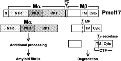
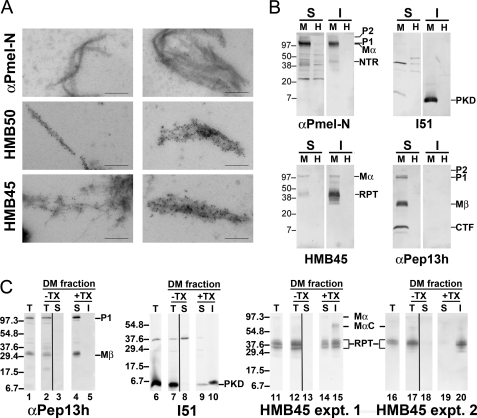
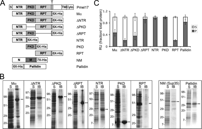
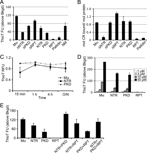
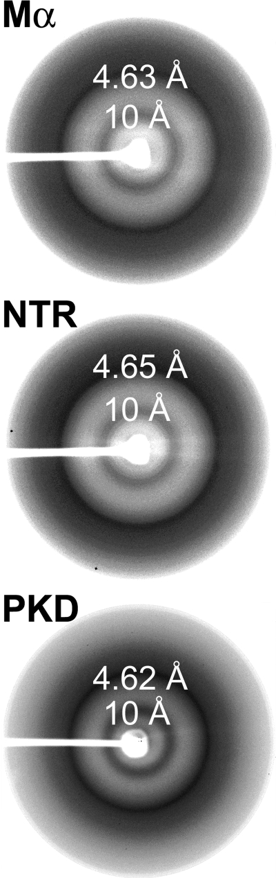
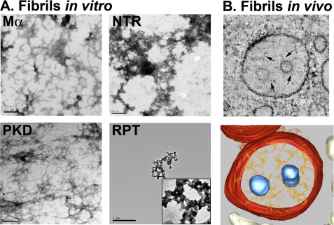
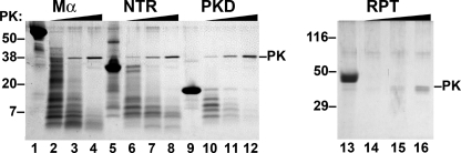
Pmel17 is a transmembrane protein that mediates the early steps in the formation of melanosomes, the subcellular organelles of melanocytes in which melanin pigments are synthesized and stored. In melanosome precursor organelles, proteolytic fragments of Pmel17 form insoluble, amyloid-like fibrils upon which melanins are deposited during melanosome maturation. The mechanism(s) by which Pmel17 becomes competent to form amyloid are not fully understood. To better understand how amyloid formation is regulated, we have defined the domains within Pmel17 that promote fibril formation in vitro. Using purified recombinant fragments of Pmel17, we show that two regions, an N-terminal domain of unknown structure and a downstream domain with homology to a polycystic kidney disease-1 repeat, efficiently form amyloid in vitro. Analyses of fibrils formed in melanocytes confirm that the polycystic kidney disease-1 domain forms at least part of the physiological amyloid core. Interestingly, this same domain is also required for the intracellular trafficking of Pmel17 to multivesicular compartments within which fibrils begin to form. Although a domain of imperfect repeats (RPT) is required for fibril formation in vivo and is a component of fibrils in melanosomes, RPT is not necessary for fibril formation in vitro and in isolation is unable to adopt an amyloid fold in a physiologically relevant time frame. These data define the structural core of Pmel17 amyloid, imply that the RPT domain plays a regulatory role in timing amyloid conversion, and suggest that fibril formation might be physically linked with multivesicular body sorting.


Figures

FIGURE 1.
FIGURE 1.
Schematic representation of Pmel17 proteolytic processing. Shown is a scheme for the primary structure of human Pmel17 and its component domains. Full-length Pmel17 is cleaved first by furin or a related proprotein convertase (PC) into Mα and Mβ fragments. Mα is incorporated into amyloid fibrils and is further proteolytically processed during fibril maturation. Mβ is cleaved by a metalloproteinase (MP), and the resulting C-terminal fragment (CTF) is a target for γ-secretase cleavage, likely facilitating the degradation of Mβ-derived fragments in lysosomes and/or proteasomes. S, signal sequence; NTR, N-terminal region; PKD, polycystic kidney disease protein homology domain; RPT, repeat domain; TM, transmembrane domain; Cyto, cytoplasmic domain.
FIGURE 2.
FIGURE 2.
Melanosome-derived fibrils are enriched in PKD and RPT domain-containing fragments. A, dense membrane fraction from MNT-1 melanoma cell homogenates was solubilized with Triton X-100, and the insoluble fraction, enriched in melanin and melanosome fibrils, was labeled with the indicated antibodies and protein A-conjugated gold particles, and then analyzed by electron microscopy. Note that both thin immature fibrils and mature fibril sheets are recognized by antibodies HMB50 (to the PKD domain) and HMB45 (to the RPT domain), but not αPmel-N (to the N-terminal peptide), indicating that they are enriched in PKD and RPT-containing fragments but lack the N terminus. Scale bars, 200 nm. B, human MNT-1 melanoma (M) and nonmelanocytic HeLa (H) cells were lysed with 1% Triton X-100 and fractionated into Triton X-100-soluble (S) and -insoluble (I) cell fractions, the latter enriched in melanosome fibrils. Fractions were subjected to SDS-PAGE followed by immunoblotting using antibodies raised against the NTR (αPmel-N), PKD (I51), RPT (HMB45), and the cytoplasmic (αPep13h) domains of Pmel17. Note that the fragments reactive with antibodies to the RPT and PKD domains in the insoluble fractions of MNT-1 cells migrate with different molecular weights, indicating that these domains are proteolytically separated within Triton X-100-insoluble fibrils. C, MNT-1 melanoma cell homogenates (T for total; lanes 1, 6, 11, and 16) were fractionated by differential sedimentation on a 2 m sucrose cushion, and a dense membrane (DM) fraction was isolated as in A (T for total; lanes 2, 7, 12, and 17). Membranes in this fraction were collected by centrifugation at 100,000 × g for 1 h; supernatants (S) were also collected (lanes 3, 8, 13, and 18). The membranes were then treated with 1% Triton X-100 (TX), and the detergent-soluble (S; lanes 4, 9, 14, and 19) and -insoluble (I; lanes 5, 10, 15, and 20) fractions were separated by sedimentation at 20,000 × g for 20 min. Equal cell equivalents of each fraction were further fractionated by SDS-PAGE and analyzed by immunoblotting with the indicated antibodies. For HMB45 immunoblots, results are shown from two different experiments, each of which was representative of at least two separate repetitions.
FIGURE 3.
FIGURE 3.
Solubility of recombinant Pmel17 His-tagged constructs. A, schematic diagram of full-length Pmel17 and C-terminally His-tagged recombinant lumenal domain fragments. Δ denotes that the indicated domain has been deleted from full-length Mα. His6, hexahistidine tag on recombinant proteins. Also shown is the His7-tagged prion-forming subdomain (NM) of the yeast prion protein Sup35, used as a positive amyloid control, and the His6-tagged full-length Pallidin, used as a negative control. B, partitioning into soluble and insoluble bacterial fractions. BL21 E. coli expressing the different proteins indicated in A were harvested and processed as described under “Experimental Procedures.” The soluble (S) and insoluble IB fractions were separated by SDS-PAGE and analyzed by Coomassie Blue staining. Asterisks denote the position of the induced protein, and the migration of molecular weight standards is indicated to the left of each pair of lanes. Note that all of the Pmel17-derived proteins are found predominantly in the IB fraction with the exception of the RPT, which is found predominantly in the soluble fraction. C, sedimentation. Each of the recombinant proteins was solubilized from inclusion bodies in guanidine HCl and affinity-purified by His-bind chromatography. Affinity-purified protein was diluted out of the denaturant into physiological buffer and allowed to refold overnight with agitation at 37 °C. Aliquots were fractionated into a soluble supernatant (S) and insoluble pellet (P) by centrifugation at 100,000 × g for 1 h at 4 °C. Total, supernatant, and pellet fractions were analyzed by SDS-PAGE, stained with Coomassie Blue, and image scanned; the relative amount of protein in each fraction, assessed as signal intensity, was determined using ImageQuant software. The mean fraction of protein in the supernatant and pellet fractions relative to the total is plotted ± S.D. n = 3 for Pallidin, n ≥ 4 all others. RU, relative unit.
FIGURE 4.
FIGURE 4.
Amyloid dye binding properties of the NTR and PKD domains, but not the RPT domain, resemble those of Mα. A, ThioT fluorescence analysis of Mα subdomains. Affinity-purified proteins in denaturant were diluted into physiological assay buffer and incubated for 16 h at 37 °C with agitation to initiate refolding and/or fibrillogenesis. Aliquots were combined with ThioT, and fluorescence emission at 490 nm was measured upon excitation at 440 nm. Columns represent the mean fluorescent units (FU) above background (ThioT alone; average value, 20 fluorescent units) ± S.E. from at least three experiments. B, Congo red binding of Mα subdomains. Renatured proteins were prepared as in A and then combined with Congo red and analyzed by light spectroscopy. Plotted are the moles of CR bound/mol of protein, as determined according to Ref. . Bars represent mean ± S.E. from at least three experiments. C, time dependence of ThioT binding for Mα subdomains. Solubilized proteins were diluted out of denaturant and incubated for the indicated times, after which ThioT was added, and fluorescence emission was measured. ThioT fluorescence intensity was normalized relative to the maximum fluorescence intensity observed. Bars represent mean ± S.E. from at least two experiments done in triplicate. Values for each of the proteins in 8 m urea (“time 0”) were negligible relative to ThioT alone. RFU, relative fluorescent units. D, protein concentration dependence on ThioT binding and fluorescence. Increasing concentrations (as indicated) of protein prepared as in A were combined with ThioT, and fluorescence emission was measured and plotted in the bar graph. Bars represent mean ± S.D. from a representative experiment. E, ThioT fluorescence of Mα subdomains alone or in combination. Solubilized proteins were diluted out of denaturant and incubated at 37 °C overnight either alone or in combination as indicated (10 μm final concentration of each protein). ThioT was added at the end of the incubation, and fluorescence emission at 490 nm was measured. Columns represent ThioT fluorescence above background from a representative experiment performed in triplicate ± S.D. Note that the signal from each combination is roughly equivalent to the sum of the signals from each component, suggesting lack of significant synergy.
FIGURE 5.
FIGURE 5.
Amyloid-like x-ray diffraction patterns of the NTR and PKD domains. Resolubilized proteins in denaturant were dialyzed against deionized water, lyophilized, and analyzed by x-ray diffraction. The diffraction patterns of Mα, NTR, and PKD are shown. Note the reflections at 4.6 and 10 Å representing the regular spacing between strands within a β-sheet and between β-sheets, respectively.
FIGURE 6.
FIGURE 6.
Refolded PKD domain has fibrillar morphology, and refolded Mα resembles melanosome fibrils. A, affinity-purified proteins, as indicated, were renatured by dilution out of the denaturant into physiological assay buffer and incubated overnight at 37 °C with agitation. Samples were then centrifuged at 100,000 × g for 1 h at 4 °C, and pellets were resuspended in a small volume of assay buffer and mounted directly on coated grids, stained with uranyl acetate, and visualized by electron microscopy. Note the branched fibrillar structures apparent in the sample containing Mα and the long fibrillar structures in samples containing the PKD, whereas the NTR appears as aggregates. Fields containing the RPT domain were difficult to find; an isolated RPT aggregate is shown at low magnification (scale bar 1 μm as compared with 0.2 μm for the others) and magnified ×5 in the inset. B, electron tomography of early stage melanosomes. MNT-1 melanoma cells preserved by high pressure freezing were analyzed by electron tomography. Top, a slice from a single tomographic reconstruction showing branched fibrils emerging from internal membrane vesicles of a multivesicular endosome. Bottom, three-dimensional model of the same tomographic reconstruction. Note the similar branched morphology of the Mα fibrils formed in vitro (A, top left panel) and the protofibrils observed in cells (B, bottom panel).
FIGURE 7.
FIGURE 7.
NTR and PKD domains, but not the RPT, are resistant to proteinase digestion. Renatured proteins were treated with increasing concentrations (1, 3.33, and 10 μg/ml) of proteinase K (PK) for 30 min at 37 °C with agitation; digestion products were fractionated by SDS-PAGE and visualized by Coomassie Blue staining. The band corresponding to proteinase K is indicated to the right, and migration of molecular weight standards is indicated to the left. Note the absence of protease-resistant fragments of the RPT domain even at the lowest proteinase K concentration but the presence of resistant fragments for all other domains.

References

    1. Berson J. F., Harper D. C., Tenza D., Raposo G., Marks M. S. (2001) Mol. Biol. Cell 12, 3451–3464 - PMC - PubMed
    1. Theos A. C., Truschel S. T., Raposo G., Marks M. S. (2005) Pigment Cell Res. 18, 322–336 - PMC - PubMed
    1. Chakraborty A. K., Platt J. T., Kim K. K., Kwon B. S., Bennett D. C., Pawelek J. M. (1996) Eur. J. Biochem. 236, 180–188 - PubMed
    1. Fowler D. M., Koulov A. V., Alory-Jost C., Marks M. S., Balch W. E., Kelly J. W. (2006) PLoS Biol. 4, e6. - PMC - PubMed
    1. Brunberg E., Andersson L., Cothran G., Sandberg K., Mikko S., Lindgren G. (2006) BMC Genet. 7, 46. - PMC - PubMed

Publication types

MeSH terms

LinkOut - more resources