. 2010 Jan;30(1):220-30.
doi: 10.1128/MCB.00552-09.

Site-specific phosphorylation induces functionally active conformation in the intrinsically disordered N-terminal activation function (AF1) domain of the glucocorticoid receptor

Affiliations

Site-specific phosphorylation induces functionally active conformation in the intrinsically disordered N-terminal activation function (AF1) domain of the glucocorticoid receptor

Anna M S Garza et al. Mol Cell Biol. 2010 Jan.

Abstract

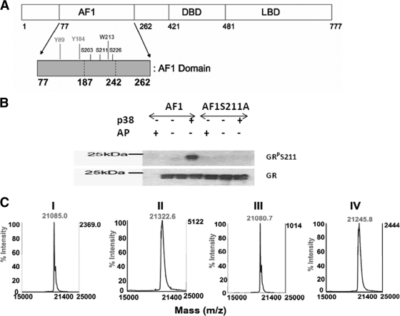
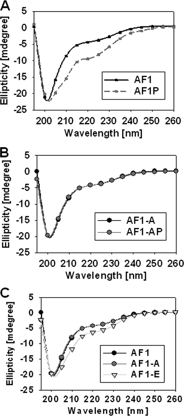
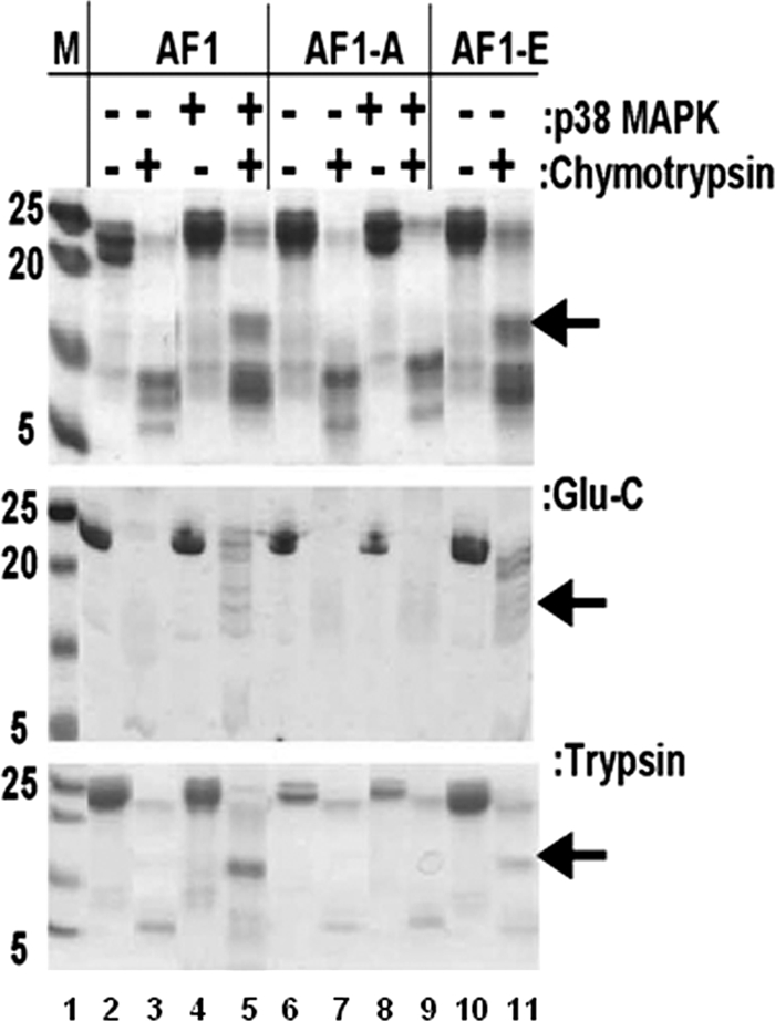
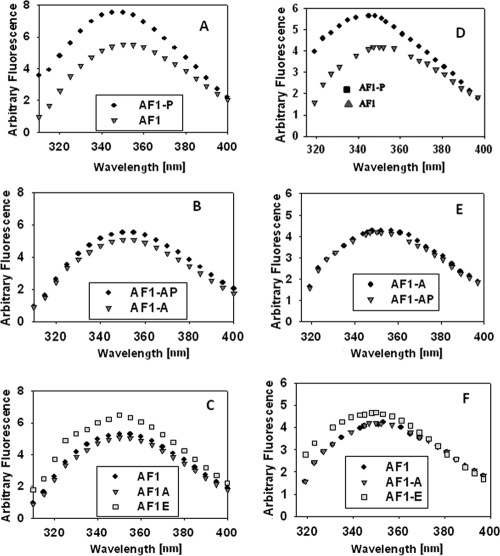
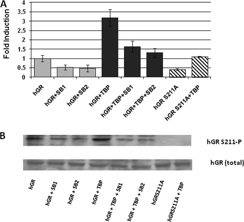
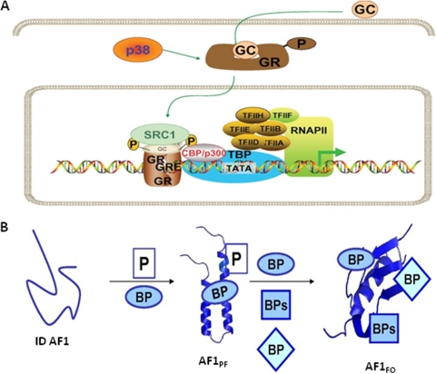
Intrinsically disordered (ID) regions are disproportionately higher in cell signaling proteins and are predicted to have much larger frequency of phosphorylation sites than ordered regions, suggesting an important role in their regulatory capacity. In this study, we show that AF1, an ID activation domain of the glucocorticoid receptor (GR), adopts a functionally folded conformation due to its site-specific phosphorylation by p38 mitogen-activated protein kinase, which is involved in apoptotic and gene-inductive events initiated by the GR. Further, we show that site-specific phosphorylation-induced secondary and tertiary structure formation specifically facilitates AF1's interaction with critical coregulatory proteins and subsequently its transcriptional activity. These data demonstrate a mechanism through which ID activation domain of the steroid receptors and other similar transcription factors may adopt a functionally active conformation under physiological conditions.


Figures

FIG. 1.
FIG. 1.
p38 phosphorylates conserved Ser residues in GR's AF1, as shown by S211-phospho-specific antibody and MALDI-TOF MS analysis. (A) Topological diagram of the hGR with expended AF1 domain containing conserved phosphorylation sites. (B) AF1 but not AF1-S211A (AF1-A) is phosphorylated at site S211. In vitro phosphorylation of AF1 and AF1-S211A was performed using p38 MAPK. AF1 or AF1-S211A and active p38 MAPK or calf alkaline phosphatase (AP) were incubated, followed by SDS-PAGE and by immunoblot analysis with GR-P S211- and GR-specific antibodies. (C) Recombinant AF1 and AF1-S211A were phosphorylated in vitro as described above, and the phosphorylation status was confirmed by MALDI MS. Subpanels: I, AF1 without p38 MAPK; II, AF1 phosphorylated with p38 MAPK; III, AF1-S211A without p38 MAPK; IV, AF1-S211A with p38 MAPK. The difference in mass between panels I and II and between panels III and IV is equivalent to three and two phospho groups, respectively.
FIG. 2.
FIG. 2.
Site-specific phosphorylation induces secondary structure in ID AF1 domain. (A to C) Far-UV CD spectra of recombinant AF1 and AF1-S211A with or without p38 MAPK and AF1-S211E. AF1, unphosphorylated AF1; AF1-P, phosphorylated; AF1-A, AF1S211A mutant; AF1-E, AF1S211E mutant. Each spectrum represents an average of five spectra recorded, corrected for the contribution of the buffer, and smoothed.
FIG. 3.
FIG. 3.
Phosphorylated AF1 resists partial proteolysis. A Coomassie blue-stained SDS-PAGE gel showing limited proteolytic digestion of purified AF1 and AF1-S211A with (+) or without (−) p38 MAPK and AF1-S211E is shown. AF1-A, AF1S211A mutant; AF1-E, AF1S211E mutant. Experiments were performed with sequencing-grade trypsin, chymotrypsin, or Endo Glu-C. Experiments were repeated at least five times with similar results.
FIG. 4.
FIG. 4.
Site-specific phosphorylation induces tertiary structure in ID AF1 domain. (A to F) Fluorescence emission spectra of AF1 and AF1-S211A with or without p38 MAPK and AF1-S211E recorded at a 278-nm (A to C) or 295-nm (D to F) excitation wavelength. AF1, unphosphorylated AF1; AF1-P, phosphorylated; AF1-A, AF1S211A mutant; AF1-E, AF1S211E mutant. Each spectrum was recorded in the emission wavelength range of 300 to 400 nm, and the result of one representative spectrum of at least three independent experiments is presented, corrected for the contribution of the buffer, and smoothed.
FIG. 5.
FIG. 5.
Phosphorylation-induced conformational changes facilitate interactions of AF1 with critical coregulatory proteins from HeLa nuclear extracts. Immunoprecipitation (IP) with indicated antibodies was carried out with HeLa cell nuclear extracts with AF1. Shown are representative immunoblots (IB) of at least three independent experiments. Graphs show the results of densitometric analysis of three experiments. The results are expressed as means ± the standard deviation. Lanes: 1, control AF1; 2, unphosphorylated AF1; 3, phosphorylated AF1; 4, unphosphorylated AF1-S211A; 5, phosphorylated AF1-S211A.
FIG. 6.
FIG. 6.
Phosphorylation-induced conformational changes facilitate interactions of AF1 with critical coregulatory proteins in CV-1 cell as assessed by FRET analyses. Representative same-cell images in the donor (CFP) channel before and after PB and the YFP channel are shown postbleaching to demonstrate the bleach efficiency. The upper panel graphs show the average FRET efficiency in each case, whereas the lower panel represents one such image in each case. (A) Controls receiving fluorescent proteins (CFP-YFP) without TBP or GR to establish basal and maximal FRET efficiency under the experimental conditions. Graph lanes: 1, CFP-empty + YFP-empty negative control; 2, CFP-YFP fusion constructs positive control. (B to D) Interactions between GR500 and TBP (B), CBP (C), or SRC-1 (D). 1, CFP-empty + YFP-TBP; 2, CFP-GR500 + YFP-empty; 3, CFP-GR500 + YFP-TBP, CBP, or SRC-1; 4, CFP-GR500-S211A + YFP-empty; 5, CFP-GR500-S211A + YFP-TBP, CBP, or SRC-1; 6, CFP-GR500-S211E + YFP-empty; 7, CFP-GR500-S211E + YFP-TBP, CBP, or SRC-1. Experiments were carried out three independent times and analyzed, and the calculated average FRET efficiencies ± the standard deviation of 15 cells were graphed for each of the conditions.
FIG. 7.
FIG. 7.
Confocal microscopic images showing that mutations (S211A or -E) within AF1 do not affect GR500's translocation to the nucleus in CV-1 cells. CV-1 cells constitutively expressing GFP-GR500, YFP-TBP, or GFP-GR500 plus YFP-TBP (row 1), GFP-GR500S211A, YFP-TBP, or GFP-GR500S211A plus YFP-TBP (row 2), or GFP-GR500S211E, YFP-TBP, or GFP-GR500S211E plus YFP-TBP (row 3) (left panels) are shown.
FIG. 8.
FIG. 8.
Phosphorylation-dependent cofactor-binding increases AF1-mediated transcriptional activity of a promoter containing 3×GRE. (A) S211A mutant inhibits GRE-mediated AF1 activity, as assessed by SEAP-based promoter-reporter assay in CV-1 cells. (B) Treatment of pharmacological inhibitors of p38 (SB203580 or SB202190) inhibits GRE-mediated GR AF1 activity in a promoter-reporter assay. SB1, SB203580; SB2, SB202190. The results are expressed as means ± the standard error. Experiments were repeated five times. The levels of significance were evaluated by a two-tailed paired Student t test, and a P value of <0.05 was considered significant. Graphs were normalized to the transfection efficiency of each construct assayed by immunoblotting with specific antibodies for GR, TBP, CBP, SRC-1, and GFP (data not shown).
FIG. 9.
FIG. 9.
Phosphorylation-dependent cofactor-binding increases GRE-mediated transcriptional activity of full-length GR. (A) S211A mutation in hGR or treatment of pharmacological inhibitors of p38 (SB203580 or SB202190) inhibits GRE-mediated hGR activity in a promoter-reporter assay. SB1, SB203580; SB2, SB202190. The results are expressed as means ± the standard error. The levels of significance were evaluated by a two-tailed paired Student t test, and a P value of <0.05 was considered significant. (B) Immunoblots showing the levels of hGR expression in transfected CV-1 cells. The upper panel shows the levels of S211 phosphorylation in hGR, and the lower panel shows the levels of total hGR in each case.
FIG. 10.
FIG. 10.
(A) Model showing the proposed effect of phosphorylation by p38 on GR-mediated transcription (based on our data and the available literature). (B) Working model of the ID AF1 domain undergoing conformational changes due to phosphorylation and/or interaction with binding partners that lead to interaction with one or more binding partners adapting functionally active ordered conformation. AF1PF, partially folded; AF1FO, fully folded AF1.

References

    1. Andrade, M.A., P. Chacón, J. J. Merelo, and F. Morán. 1993. Evaluation of secondary structure of proteins from UV circular dichroism using an unsupervised learning neural network. Protein Eng. 6:383-390. - PubMed
    1. Bai, Y., and V. Giguére. 2003. Isoform-selective interactions between estrogen receptors and steroid receptor coactivators promoted by estradiol and ErbB-2 signaling in living cells. Mol. Endocrinol. 17:589-599. - PubMed
    1. Baldwin, R. L., and G. D. Rose. 1999. Is protein folding hierarchic? II. Folding intermediates and transition states. Trends Biochem. Sci. 24:77-83. - PubMed
    1. Baskakov, I. V., R. Kumar, G. Srinivasan, Y. Ji, D. W. Bolen, and E. B. Thompson. 1999. Trimethylamine N-oxide-induced cooperative folding of an intrinsically unfolded transcription-activating fragment of human glucocorticoid receptor. J. Biol. Chem. 274:10693-10696. - PubMed
    1. Blind, R. D., and M. J. Garabedian. 2008. Differential recruitment of glucocorticoid receptor phospho-isoforms to glucocorticoid-induced genes. J. Steroid Biochem. Mol. Biol. 109:150-157. - PMC - PubMed

Publication types

MeSH terms

Substances