. 2010 Jan;30(1):98-105.
doi: 10.1128/MCB.01155-09.

Functional cooperation of the proapoptotic Bcl2 family proteins Bmf and Bim in vivo

Affiliations

Functional cooperation of the proapoptotic Bcl2 family proteins Bmf and Bim in vivo

Anette Hübner et al. Mol Cell Biol. 2010 Jan.

Abstract

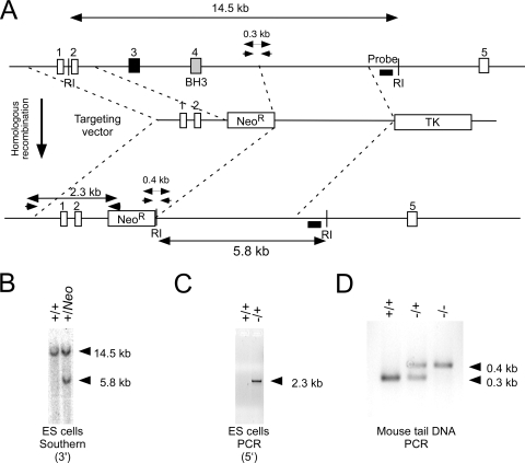
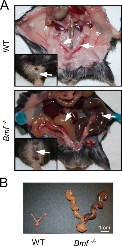
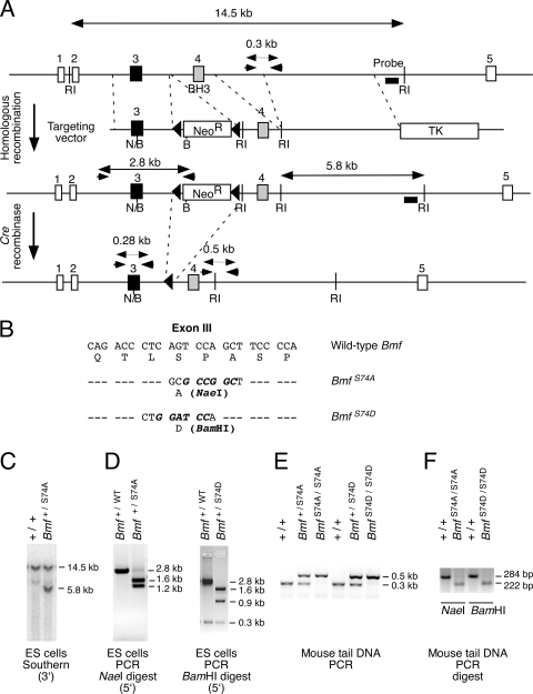
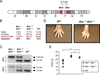
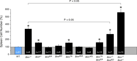
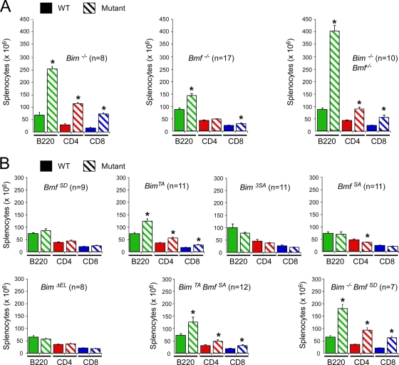
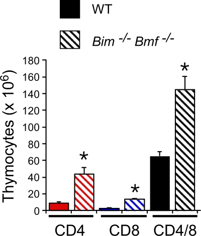
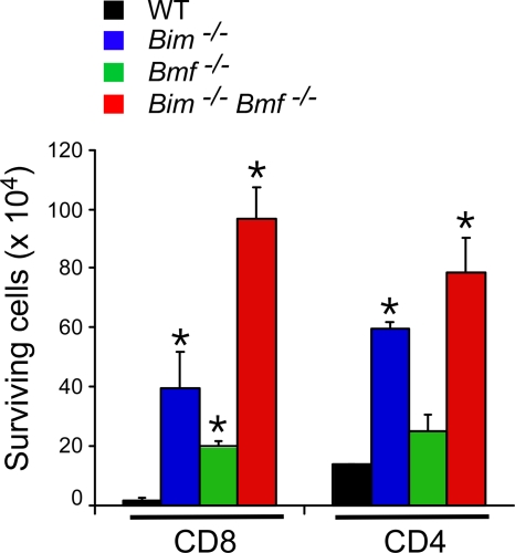
Bcl2-modifying factor (Bmf) is a member of the BH3-only group of proapoptotic proteins. To test the role of Bmf in vivo, we constructed mice with a series of mutated Bmf alleles that disrupt Bmf expression, prevent Bmf phosphorylation by the c-Jun NH(2)-terminal kinase (JNK) on Ser(74), or mimic Bmf phosphorylation on Ser(74). We report that the loss of Bmf causes defects in uterovaginal development, including an imperforate vagina and hydrometrocolpos. We also show that the phosphorylation of Bmf on Ser(74) can contribute to a moderate increase in levels of Bmf activity. Studies of compound mutants with the related gene Bim demonstrated that Bim and Bmf exhibit partially redundant functions in vivo. Thus, developmental ablation of interdigital webbing on mouse paws and normal lymphocyte homeostasis require the cooperative activity of Bim and Bmf.


Figures

FIG. 1.
FIG. 1.
Disruption of the murine Bmf gene. (A) Strategy for construction of Bmf/ mice. The structure of the Bmf genomic locus and the targeting vector are illustrated. EcoRI (RI) restriction sites are indicated. Homologous recombination causes the replacement of Bmf exons 3 and 4 with a Neor cassette. The PCR amplimers to confirm 5′ integration and the Southern blot probe to confirm 3′ integration of the targeting vector are indicated. The PCR amplimers employed for genotype analysis are also illustrated. (B) Southern blot analysis of genomic DNA confirms 3′ integration of the targeting vector in ES cells. (C) PCR analysis of genomic DNA confirms the 5′ integration of the targeting vector in ES cells. (D) Genomic DNA isolated from wild-type, Bmf/+, and Bmf/ mice was examined by PCR analysis.
FIG. 2.
FIG. 2.
Uterovaginal abnormalities in Bmf/ mice. (A) (Upper panel) Reproductive tract of a wild-type (WT) female mouse with a normal uterus and fallopian tubes (arrow) and vagina (insert). (Lower panel) Reproductive tract of a Bmf/ female mouse with an imperforate vagina (insert) and hydrometrocolpos (arrow). (B) Dissected female genital tracts of wild-type and Bmf/ female mice.
FIG. 3.
FIG. 3.
Construction of mice with disrupted Bmf phosphorylation. (A) Strategy for construction of mice with mutation of the Bmf phosphorylation site at Ser74. The structure of the Bmf genomic locus and the targeting vectors are illustrated. Homologous recombination causes the replacement of Bmf exon 3 with a mutated form of exon 3 together with the insertion of a floxed Neor resistance cassette. The PCR amplimers to confirm 5′ integration and the Southern blot probe to confirm 3′ integration of the targeting vector are indicated. The PCR amplimers employed for genotype analysis are also illustrated. Restriction endonuclease sites are also illustrated (BamHI [B], EcoRI [RI], and NaeI [N]). (B) The point mutations in exon 3 introduced by the targeting vectors are illustrated. (C and D) Homologous integration was confirmed by Southern blot analysis (3′ end) (C) and by PCR (5′ end) (D). The presence of S74A (NaeI) and S74D (BamHI) was detected by digestion of the 2.8-kb PCR product with the indicated restriction enzymes. (E) Mouse tail DNA was genotyped by PCR. (F) The genotype at codon 74 was examined by restriction digestion (NaeI and BamHI) of PCR products obtained using amplimers that span Bmf exon 3.
FIG. 4.
FIG. 4.
Mice with compound deficiency of Bim and Bmf. (A) The location of the Bim and Bmf genes on chromosome 2 is illustrated. These genes are present within a 9.5-Mb region of mouse chromosome 2. (B) Mice with one wild-type chromosome 2 and one Bim/Bmf mutant chromosome 2 were intercrossed. The numbers of wild-type, double-heterozygous, and double-homozygous progeny are presented as the percentage of the total progeny (n = 120). The expected Mendelian ratios of progeny are also shown. (C) Genotype analysis of a wild-type mouse and compound mutant mice (Bim/+ Bmf/+ and Bim/ Bmf/) was performed by PCR amplification of genomic DNA. (D) The webbed foot of a Bim/ Bmf/ mouse is compared with the foot of a wild-type mouse. (E) The masses of wild-type, Bim/+ Bmf/+, and Bim/Bmf / mice (aged 6 weeks) were measured. The masses of six individual mice per group are shown. The mean mass is illustrated with a horizontal line. Statistically significant differences are indicated (*, P < 0.05).
FIG. 5.
FIG. 5.
Bim and Bmf deficiency causes splenomegaly. The relative numbers of splenocytes isolated from wild-type mice (100%) and mice with homozygous defects in the Bim and/or Bmf gene are presented (means ± standard deviations). Statistically significant differences between mutant mice and wild-type mice are indicated (*, P < 0.05). The figure also shows statistically significant differences (P < 0.05) between Bim/ mice and Bim/ BmfSD/SD or Bim/ Bmf/ mice.
FIG. 6.
FIG. 6.
Bim and Bmf deficiency causes an increase in the number of B cells and T cells in the spleen. (A and B) Spleen cells were examined by flow cytometry to identify B cells and T cells using antibodies to cell surface B220, CD4, and CD8 (means ± standard deviations). Statistically significant differences between wild-type and mutant mice are indicated (*, P > 0.05).
FIG. 7.
FIG. 7.
Effect of compound mutation of Bim and Bmf on thymocytes. Thymocytes isolated from wild-type and Bim/ Bmf/ mice were examined by flow cytometry using antibodies to cell surface CD4 and CD8 (means ± standard deviations; n = 6). Statistically significant differences between wild-type and mutant cells are indicated (*, P < 0.05).
FIG. 8.
FIG. 8.
Bmf and Bim deficiency causes reduced T-cell apoptosis. Purified CD4 and CD8 T cells (1 × 10⁶ cells) were incubated in medium (4 days), and the number of viable cells was counted (means ± standard deviations; n = 5). Statistically significant differences between wild-type and mutant cells are indicated (*, P < 0.05).

References

    1. Bouillet, P., D. Metcalf, D. C. Huang, D. M. Tarlinton, T. W. Kay, F. Kontgen, J. M. Adams, and A. Strasser. 1999. Proapoptotic Bcl-2 relative Bim required for certain apoptotic responses, leukocyte homeostasis, and to preclude autoimmunity. Science 286:1735-1738. - PubMed
    1. Cecconi, F., G. Alvarez-Bolado, B. I. Meyer, K. A. Roth, and P. Gruss. 1998. Apaf1 (CED-4 homolog) regulates programmed cell death in mammalian development. Cell 94:727-737. - PubMed
    1. Davis, R. J. 2000. Signal transduction by the JNK group of MAP kinases. Cell 103:239-252. - PubMed
    1. Diehl, S., C. W. Chow, L. Weiss, A. Palmetshofer, T. Twardzik, L. Rounds, E. Serfling, R. J. Davis, J. Anguita, and M. Rincon. 2002. Induction of NFATc2 expression by interleukin 6 promotes T helper type 2 differentiation. J. Exp. Med. 196:39-49. - PMC - PubMed
    1. Farley, N., G. Pedraza-Alva, D. Serrano-Gomez, V. Nagaleekar, A. Aronshtam, T. Krahl, T. Thornton, and M. Rincon. 2006. p38 mitogen-activated protein kinase mediates the Fas-induced mitochondrial death pathway in CD8+ T cells. Mol. Cell. Biol. 26:2118-2129. - PMC - PubMed

Publication types

MeSH terms