. 2009 Nov 3;106(44):18751-6.
doi: 10.1073/pnas.0905010106. Epub 2009 Oct 19.

An essential role for RPE-derived soluble VEGF in the maintenance of the choriocapillaris

Affiliations

An essential role for RPE-derived soluble VEGF in the maintenance of the choriocapillaris

Magali Saint-Geniez et al. Proc Natl Acad Sci U S A. .

Abstract

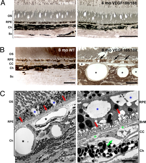
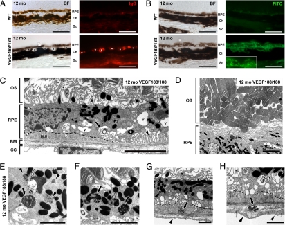
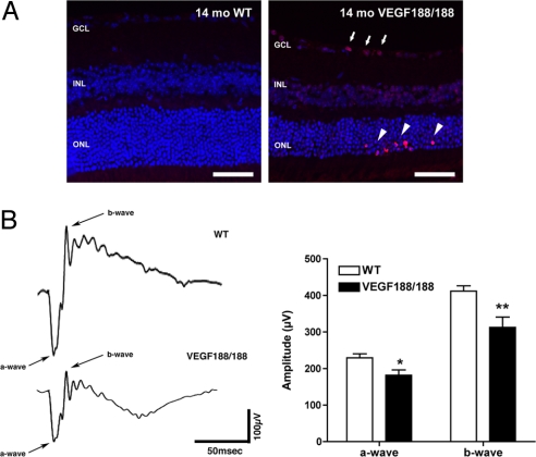
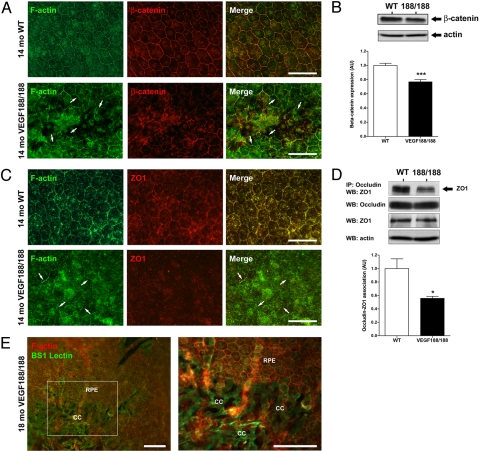
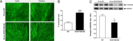
Clinical and experimental observations indicate a role for VEGF secreted by the retinal pigment epithelium (RPE) in the maintenance of the choriocapillaris (CC). VEGF in mice is produced as three isoforms, VEGF120, VEGF164, and VEGF188, that differ in their ability to bind heparan sulfate proteoglycan. RPE normally produces the more soluble isoforms, VEGF120 and VEGF164, but virtually no VEGF188, reflecting the fact that molecules secreted by the RPE must diffuse across Bruch's membrane (BrM) to reach the choriocapillaris. To determine the role of RPE-derived soluble VEGF on the choriocapillaris survival, we used mice that produce only VEGF188. VEGF188/188 mice exhibited normal choriocapillaris development. However, beginning at 7 months of age, we observed a progressive degeneration characterized by choriocapillaris atrophy, RPE and BrM abnormalities, culminating in areas of RPE loss and dramatic choroidal remodeling. Increased photoreceptor apoptosis in aged VEGF188/188 mice led to a decline in visual acuity as detected by electroretinogram (ERG). These changes are reminiscent of geographic atrophy (GA) and point to a role for RPE-derived VEGF in the maintenance of the choriocapillaris.


Conflict of interest statement

The authors declare no conflict of interest.

Figures

Fig. 1.
Fig. 1.
Abnormal choroidal vasculature and RPE defects in aged VEGF188/188 mice. (A and B) One-micrometer-thin epon sections of 4- and 8-month-old wt (right panel) and VEGF188/188 (left panel) mice were stained with para-phenylenediamine for light microscopy (A and B) or were visualized by TEM (C). (A) No changes in the organization of the retina layers were observed in 4-month-old VEGF188/188 mice. (B) An 8-month-old, VEGF188/188 mouse eye displays major abnormalities in the RPE and choroidal vessels. Larges vacuoles were observed within the RPE (arrows). The choroidal vessels appear enlarged, and the CC is replaced by discontinuous vessels wrapped by pericytes (asterisk). (C) Electron micrograph of the mid-periphery of an 8-month-old VEGF188/188 mouse retina. Note the significant enlargement of the choroidal vessels (black asterisk). RPE cells displayed large vacuoles containing membranous debris (blue asterisk). Regression of the RPE basal infoldings is associated with accumulation of materials (red arrow). Higher magnification (right) showing discontinuous collagen and elastin layers in the BrM (green asterisk) and a regressing CC vessel with an entrapped erythrocyte (green arrow). [Scale bar, 50 μm (A); scale bar, 20 μm (B); scale bar, 2 μm (C).] OS, outer segment; BrM, Bruch's membrane; CC, choriocapillaris; Ch, choroid; Sc, sclera.
Fig. 2.
Fig. 2.
Early AMD features in aged VEGF188/188 mice. (A) IgG staining (red) of the choroids of 12-month-old mice reveal the deposition of IgG in the walls of the choroidal vessel of the transgenic mice (asterisks) but not in aged-matched controls. (B) Increased lipofuscin autofluorescence was detected with an FITC filter. Multiple bright dots and clusters were evident in the RPE layer of the VEGF188/188 mice, especially in the clumped cells (see insert). (C–H) Representative electron micrographs showing RPE and BrM anomalies in 12-month-old VEGF188/188 mice. (C) Unhealthy RPE cells with reduced and disorganized basal infoldings (arrowhead) displaying larges vacuoles (V) and membranous debris (large arrow). Formation of continuous subRPE deposits is outlined by a dotted line. Note the loss of intimate association between the RPE and the OS. The OS were separated from the RPE by a large space (double-headed arrow) containing disorganized RPE apical digitations and lamellar bodies (small arrows). (D) Lower magnification showing highly disorganized OS. (E and F) Higher magnification showing lipofuscin granules (arrowheads), abnormal phagosomes containing melanin (arrows in panel E), and undigested photoreceptor outer-segments in VEGF188/188 RPE (arrow in panel F). Lipid droplets were also observed in the lipofuscin bodies (open arrows). (G and H) Basal laminar deposits were associated with dense and membranous debris in the subRPE space or in the outer layers of the BrM (arrows in panels G and H, respectively). Note the loss of fenestrations and thickening of the endothelium membrane in the CC underlying the subretinal lesions. OS, outer segment; BrM, Bruch's membrane; CC, choriocapillaris; BLD, basal laminar deposits. [Scale bar, 50 μm (A and B); scale bar, 5 μm (C and D; scale bar, 2 μm (E–H).]
Fig. 3.
Fig. 3.
Increased photoreceptor apoptosis and abnormal ERGs in VEGF188/188 mice. (A) TUNEL staining revealed a markedly increased apoptosis in the ONL of 14-month-old VEGF188/188 mice. Apoptotic photoreceptors appeared as clusters in the outermost part of the ONL layer (arrowhead) and were often associated with increased cell death in the GCL (arrows). (B) Scotopic ERG recordings of 14-month-old mice using a flash intensity of +10 dB revealed a marked reduction of both a- and b-wave amplitudes in VEGF188/188 mice (n = 14 for wt and n = 12 for VEGF188/188). GCL, ganglion cell layer; INL, inner nuclear layer; ONL, outer nuclear layer. (Scale bars, 50 μm.)
Fig. 4.
Fig. 4.
RPE dystrophy is associated with barrier function loss in VEGF188/188 mice. (A) Flat-mount preparation of 14-month-old wt mice and VEGF188/188 showed abnormal distribution of the junctional proteins in RPE layer of transgenic mice. In wt, RPE formed a regular honeycomb pattern. In VEGF188/188, the RPE organization is disturbed and atrophic revealing the choroidal vessel underneath (CC). In the RPE lesions, the gap-junction protein β-catenin has lost its characteristic membranous association (arrows). (B) Abnormal β-catenin distribution was accompanied by a reduction of protein expression showed by Western blot on 16-month-old choroid-RPE samples (n = 6). (C) Aberrant ZO1 localization was also observed in the VEGF188/188 mice. (D) No changes in ZO1 protein level were detected. Immunoprecipitation for occludin followed by ZO1 Western blotting revealed a 45% reduction in association between ZO1 and occludin (n = 3). (E) Over time, the limited RPE lesions observed at 14 months (see panels A and C) expanded significantly leading to a major loss in the integrity of the RPE layer of 18-month-old VEGF188/188 mice. [Scale bar, 50 μm (A and C); scale bar, 100 μm (E).]
Fig. 5.
Fig. 5.
CC atrophy is associated with decreased VEGFR2 phosphorylation in aged VEGF188/188 mice. (A) Choroidal vasculature of 18-month-old wt and VEGF188/188 mice was immunostained by cardiac perfusion of FITC-labeled Bandeiraea simplicifolia (BS-1) isolectin. In wt animals, flat-mounted choroids revealed a typical dense honeycomb vascular network. However, the choroidal vessels of VEGF188/188 animals appeared poorly branched and atrophied. The reduced vascular density was observed in both the central and the peripheral choroidal vasculature. (Scale bar, 200 μm.) (B) Quantification of the avascular area in the CC 15- to 16-month-old VEGF188/188 mice (n = 5) compared to aged-matched wt (n = 5) (P < 0.0001). (C) Level of VEGFR2 phosphorylation in choroid-RPE protein lysates of 16-month-old mice was quantified by Western blotting using [pY1214]-VEGFR2 specific antibody. The level of phosphorylated VEGFR2 was normalized for total VEGFR2. VEGF188/188 RPE-choroid complex displayed a significant reduction of the phosphorylation level of VEGFR2 compared to wt. (Scale bar, 100 μm.)

References

    1. Holmes DI, Zachary I. The vascular endothelial growth factor (VEGF) family: Angiogenic factors in health and disease. Genome Biol. 2005;6:209. - PMC - PubMed
    1. Olsson AK, Dimberg A, Kreuger J, Claesson-Welsh L. VEGF receptor signalling—in control of vascular function. Nat Rev Mol Cell Biol. 2006;7:359–371. - PubMed
    1. Soker S, Miao HQ, Nomi M, Takashima S, Klagsbrun M. VEGF165 mediates formation of complexes containing VEGFR-2 and neuropilin-1 that enhance VEGF165-receptor binding. J Cell Biochem. 2002;85:357–368. - PubMed
    1. Maharaj AS, Saint-Geniez M, Maldonado AE, D'Amore PA. Vascular endothelial growth factor localization in the adult. Am J Pathol. 2006;168:639–648. - PMC - PubMed
    1. Eremina V, Quaggin SE. The role of VEGF-A in glomerular development and function. Curr Opin Nephrol Hypertens. 2004;13:9–15. - PubMed

Publication types

MeSH terms

Substances

LinkOut - more resources