. 2009 Dec 18;284(51):35951-61.
doi: 10.1074/jbc.M109.048330.

Identification of SMARCAL1 as a component of the DNA damage response

Affiliations

Identification of SMARCAL1 as a component of the DNA damage response

Lisa Postow et al. J Biol Chem. .

Abstract

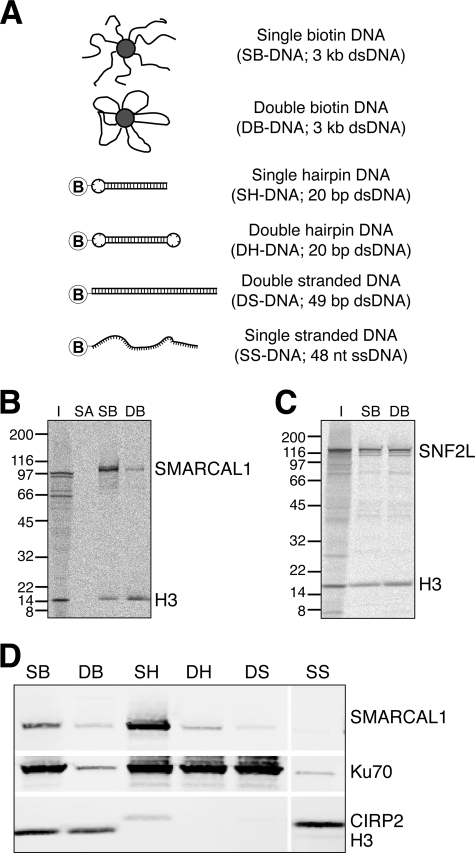
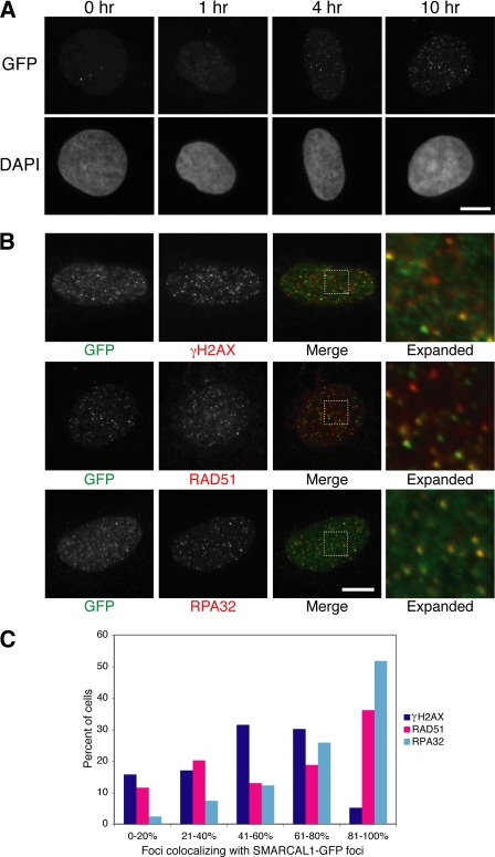
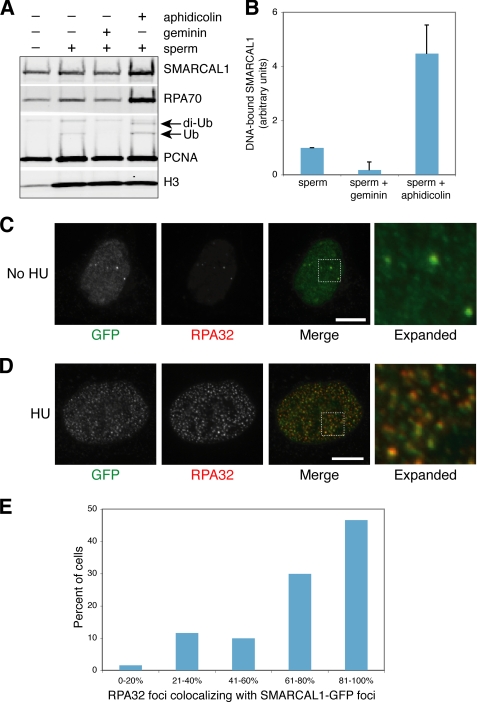
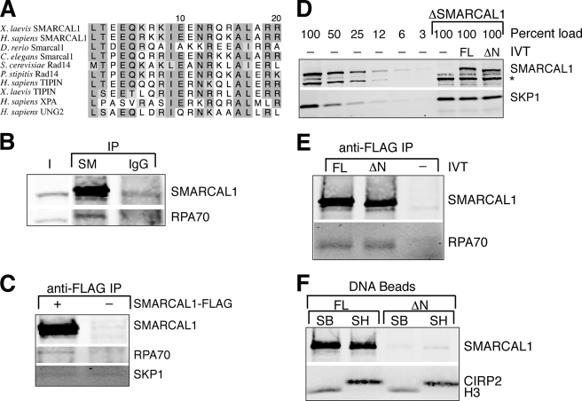
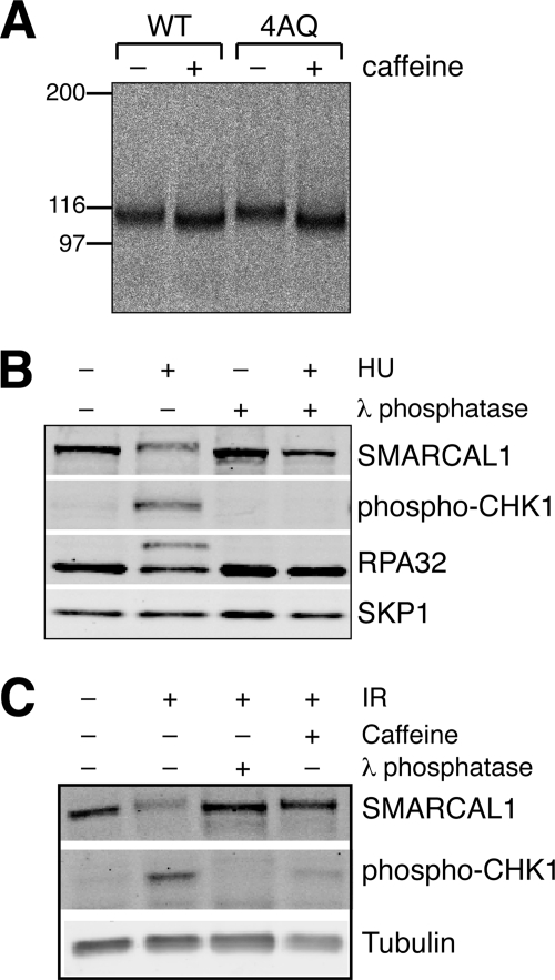
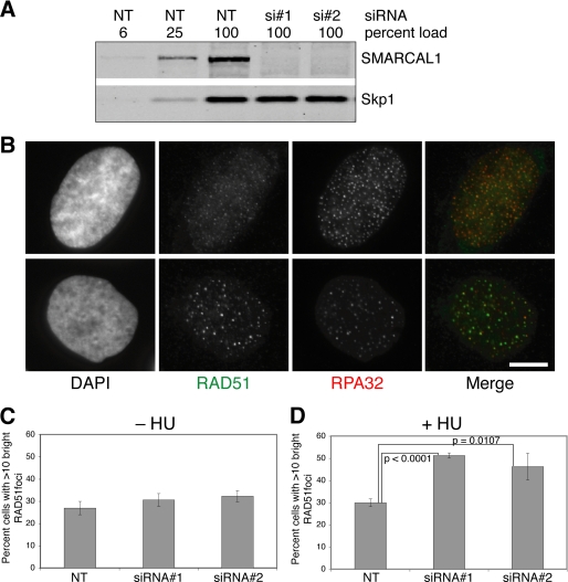
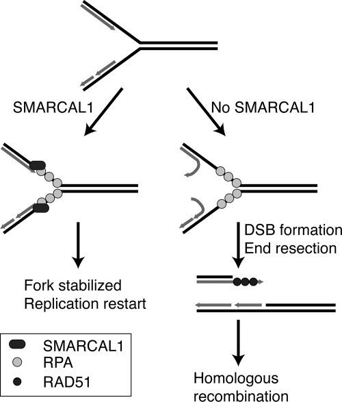
SMARCAL1 (also known as HARP) is a SWI/SNF family protein with an ATPase activity stimulated by DNA containing both single-stranded and double-stranded regions. Mutations in SMARCAL1 are associated with the disease Schimke immuno-osseous dysplasia, a multisystem autosomal recessive disorder characterized by T cell immunodeficiency, growth inhibition, and renal dysfunction. The cellular function of SMARCAL1, however, is unknown. Here, using Xenopus egg extracts and mass spectrometry, we identify SMARCAL1 as a protein recruited to double-stranded DNA breaks. SMARCAL1 binds to double-stranded breaks and stalled replication forks in both egg extract and human cells, specifically colocalizing with the single-stranded DNA binding factor RPA. In addition, SMARCAL1 interacts physically with RPA independently of DNA. SMARCAL1 is phosphorylated in a caffeine-sensitive manner in response to double-stranded breaks and stalled replication forks. It has been suggested that stalled forks can be stabilized by a mechanism involving caffeine-sensitive kinases, or they collapse and subsequently recruit Rad51 to promote homologous recombination repair. We show that depletion of SMARCAL1 from U2OS cells leads to increased frequency of RAD51 foci upon generation of stalled replication forks, indicating that fork breakdown is more prevalent in the absence of SMARCAL1. We propose that SMARCAL1 is a novel DNA damage-binding protein involved in replication fork stabilization.


Figures

FIGURE 1.
FIGURE 1.
Identification and confirmation of xSMARCAL1 as a DSB-binding protein. A, schematic representation of DNA structures used in this study. Biotinylated nucleotides were added to one or both ends of restriction enzyme-digested pBluescriptSK+ (3 kb) using the Klenow fragment of DNA polymerase I. Biotinylated DNA was then bound to streptavidin-coated magnetic beads. When one end contained biotinylated nucleotides (SB-DNA), the exposed end resembled a DSB. When both ends were biotinylated (DB-DNA), they were obscured by the bead. Additional DNA structures were constructed using biotinylated oligonucleotides. The SH-DNA structure is a 4-nucleotide biotinylated hairpin on a 20-bp stem, and the double hairpin DNA (DH-DNA) is identical except that the end is capped with a second 4-nucleotide hairpin. The double-stranded DNA (DS-DNA) is the product of annealing two 49-mer complementary oligonucleotides, one of which contains biotin at its 3′-end. Single-stranded DNA (SS-DNA) is a 48-mer that contains biotin at its 3′-end. SB-DNA and DB-DNA are shown bound to a streptavidin-coated bead. For all other DNAs, biotin is depicted by a circled B. B, following the identification of xSMARCAL1 by tandem mass spectrometry, in vitro translated xSMARCAL1 and histone H3 were added to cytostatic factor egg extract and allowed to bind to streptavidin-coated (SA), SB-DNA (SB), or DB-DNA (DB) beads, and interacting proteins were analyzed using a PhosphorImager. Although the control H3 binds equally well to SB-DNA and DB-DNA beads, xSMARCAL1 has a clear preference for SB-DNA beads. C, in contrast to the preference of xSMARCAL1 for SB-DNA beads, the chromatin remodeler xSNF2L showed no preference. D, SB-DNA (SB), DB-DNA (DB), SH-DNA (SH), double hairpin DNA (DH), double-stranded DNA (DS), and single-stranded DNA (SS) beads were incubated in egg extract for 30 min, after which associated proteins were isolated and analyzed by immunoblot. To control for DNA structural elements, immunoblots were probed with antibodies against xKu70, xCIRP2, and H3.
FIGURE 2.
FIGURE 2.
hSMARCAL1 forms damage-dependent foci that colocalize with hRPA. A, U2OS cells expressing hSMARCAL1-GFP were treated with 10 Gy of γ-irradiation, fixed at the indicated time points, and analyzed by immunofluorescence using a mouse anti-GFP antibody. Characteristic cells containing foci are shown. Bar, 10 μm. B, cells expressing hSMARCAL1-GFP were treated with 10 Gy of irradiation, fixed after 10 h, and analyzed by immunofluorescence using mouse anti-γH2AX and rabbit anti-GFP, rabbit anti-hRAD51 and mouse anti-GFP, or mouse anti-hRPA32 and rabbit anti-GFP antibodies. hSMARCAL1-GFP and hRPA32 colocalize, as seen by an expansion of the boxed regions. Bar, 10 μm. C, quantification of colocalization of DNA damage marker foci with hSMARCAL1-GFP foci. Among all detectable γH2AX (n = 74 cells), hRAD51 (n = 69 cells), and hRPA32 (n = 81 cells) foci, the fraction (percentage) of those that colocalized with hSMARCAL1-GFP was measured for each cell. Distribution of the colocalization frequency is shown.
FIGURE 3.
FIGURE 3.
SMARCAL1 colocalizes with RPA at stalled replication forks. A, sperm nuclei (4000/μl) were incubated with interphase egg extract to allow for chromosomal replication. Geminin was added before the addition of chromatin to inhibit replication initiation, and aphidicolin was added 15 min after the addition of chromatin to stall replication forks, as indicated. Chromatin was enriched by spinning through a 30% sucrose cushion after a 1-h total incubation. Co-sedimenting proteins were analyzed by immunoblot. As a control, extract was incubated in the absence of sperm chromatin but otherwise treated identically. Ub, monoubiquitylated PCNA, resulting from DNA replication; di-Ub, diubiquitylated PCNA, resulting from stalled replication forks. B, quantification of chromatin-binding xSMARCAL1 from three independent spin-down experiments. Error bars, S.D. C, normally growing U2OS cells stably expressing hSMARCAL1-GFP were co-stained with antibodies against GFP and hRPA32. An expanded region demonstrates the colocalization. Bar, 10 μm. D, U2OS cells stably expressing hSMARCAL1-GFP were treated with 4 mm HU for 24 h and then fixed and co-stained with anti-hRPA32 and anti-GFP antibodies. Bar, 10 μm. E, quantification of colocalization of hRPA32 foci with hSMARCAL1-GFP foci. Among all detectable hRPA32 foci, the fraction (percentage) of those that colocalized with hSMARCAL1-GFP was measured for each cell. Distribution of the colocalization frequency is shown (n = 60 cells).
FIGURE 4.
FIGURE 4.
xSMARCAL1 interacts with Xenopus RPA in egg extracts. A, alignment of the N terminus of SMARCAL1 with similar sequences in other proteins. The UNG2 and XPA sequences are known to interact with the C-terminal domain of RPA32 (27). B, xSMARCAL1 was immunoprecipitated using an anti-xSMARCAL1 antibody or rabbit IgG as a control, and the isolated proteins were analyzed by immunoblot. I, input; SM, anti-xSMARCAL1 antibody. C, xSMARCAL1-FLAG translated in reticulocyte lysate or a control reticulocyte lysate reaction was mixed with egg extract and incubated with anti-FLAG antibodies. Isolated proteins were analyzed by immunoblot. D, xSMARCAL1 was depleted out of extract, and reticulocyte lysate-expressed xSMARCAL1-FLAG or ΔN-xSMARCAL1-FLAG was added. The asterisk marks a nonspecific band that interacts with the anti-xSMARCAL1 antibody. IVT, in vitro translated. These extracts were used for the experiments shown in E and F. E, FLAG-tagged full-length (FL) or truncated (ΔN) xSMARCAL1 were immunoprecipitated using an anti-FLAG antibody, and isolated proteins were analyzed by immunoblot. F, xSMARCAL1-FLAG or ΔN-xSMARCAL1-FLAG was allowed to bind to SB-DNA or SH-DNA beads, and DNA-bound proteins were analyzed by immunoblot. Histone H3 and the ssDNA-binding protein xCIRP2 were used as DNA isolation controls for SB-DNA and SH-DNA beads, respectively. IP, immunoprecipitation.
FIGURE 5.
FIGURE 5.
xSMARCAL1 and hSMARCAL1 are phosphorylated in response to the DNA damage checkpoint. A, wild type xSMARCAL1 (WT) or a mutant version in which all four (S/T)Q sites were mutated to AQ (4AQ), was expressed in reticulocyte lysate, mixed with egg extract, and allowed to bind to SB-DNA beads in the absence or presence of caffeine, and bound proteins were visualized using a PhosphorImager. B, U2OS cells were grown in the presence or absence of 4 mm HU for 24 h, and lysates were treated with λ-phosphatase, as indicated. C, hSMARCAL1 mobility was analyzed after U2OS cells grown in the presence or absence of caffeine were treated with or without 10 Gy of irradiation (IR) and collected 24 h later. Lysates were treated with λ-phosphatase, as indicated.
FIGURE 6.
FIGURE 6.
hSMARCAL1-depleted cells have a higher frequency of hRAD51 foci following HU treatment. A, U2OS cells were treated with control non-targeting siRNAs (NT) or siRNAs targeting two distinct regions of the hSMARCAL1 gene (si#1 and si#2), and depletion was tested by immunoblot. More than 94% of the endogenous SMARCAL1 was depleted by either siRNA. B, examples of two types of cells following HU treatment. U2OS cells were grown in the presence of 4 mm HU for 24 h, fixed, and probed with anti-hRAD51 and anti-hRPA32 antibodies. Representative cells displaying dim hRAD51 foci with hundreds of bright hRPA32 foci (top) and bright hRAD51 foci with fewer hRPA32 foci (bottom) are shown. The cells shown were treated with siRNA 3. Bar, 10 μm. C, U2OS cells were treated with nontargeting siRNAs or siRNAs targeting hSMARCAL1 (siRNA 1 and 2) and analyzed by immunofluorescence to determine the percentage of nuclei with greater than 10 bright hRAD51 foci. The average of three independent experiments is shown, with S.D. n > 500 cells for each sample. D, cells were treated with siRNAs and grown in 4 mm HU for 24 h, after which cells with bright hRAD51 foci were counted. Significantly more hRAD51-positive nuclei were present in hSMARCAL1-depleted cells than in control cells. The average of three independent experiments is shown, with S.D. n > 500 cells for each sample.
FIGURE 7.
FIGURE 7.
A model for SMARCAL1 function at stalled replication forks. SMARCAL1 binds at the single strand/double strand junction in the replicating arm, partially through an interaction between its N terminus and the C-terminal domain of RPA32. In an ATP-dependent manner, it stabilizes base pairing between the daughter and parent strands. In the absence of SMARCAL1, incomplete daughter strands are allowed to peel off their parent strands, leading to aberrant DNA structures and replication fork instability. Following a break at the fork, the end is resected, RAD51 is loaded, and the DNA enters the homologous recombination pathway.

References

    1. Hoeijmakers J. H. (2001) Nature 411, 366–374 - PubMed
    1. Fanning E., Klimovich V., Nager A. R. (2006) Nucleic Acids Res. 34, 4126–4137 - PMC - PubMed
    1. Mimitou E. P., Symington L. S. (2009) Trends Biochem. Sci. 34, 264–272 - PubMed
    1. Zou L., Elledge S. J. (2003) Science 300, 1542–1548 - PubMed
    1. Byun T. S., Pacek M., Yee M. C., Walter J. C., Cimprich K. A. (2005) Genes Dev. 19, 1040–1052 - PMC - PubMed

Publication types