. 2010 Feb 1;19(3):397-410.
doi: 10.1093/hmg/ddp483. Epub 2009 Oct 20.

Tsc/mTORC1 signaling in oocytes governs the quiescence and activation of primordial follicles

Affiliations

Tsc/mTORC1 signaling in oocytes governs the quiescence and activation of primordial follicles

Deepak Adhikari et al. Hum Mol Genet. .

Abstract

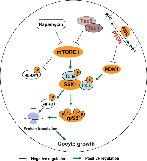
To maintain the female reproductive lifespan, the majority of ovarian primordial follicles are preserved in a quiescent state in order to provide ova for later reproductive life. However, the molecular mechanism that maintains the long quiescence of primordial follicles is poorly understood. Here we provide genetic evidence to show that the tumor suppressor tuberous sclerosis complex 1 (Tsc1), which negatively regulates mammalian target of rapamycin complex 1 (mTORC1), functions in oocytes to maintain the quiescence of primordial follicles. In mutant mice lacking the Tsc1 gene in oocytes, the entire pool of primordial follicles is activated prematurely due to elevated mTORC1 activity in the oocyte, ending up with follicular depletion in early adulthood and causing premature ovarian failure (POF). We further show that maintenance of the quiescence of primordial follicles requires synergistic, collaborative functioning of both Tsc and PTEN (phosphatase and tensin homolog deleted on chromosome 10) and that these two molecules suppress follicular activation through distinct ways. Our results suggest that Tsc/mTORC1 signaling and PTEN/PI3K (phosphatidylinositol 3 kinase) signaling synergistically regulate the dormancy and activation of primordial follicles, and together ensure the proper length of female reproductive life. Deregulation of these signaling pathways in oocytes results in pathological conditions of the ovary, including POF and infertility.


Figures

Figure 1.
Figure 1.
Oocyte-specific deletion of Tsc1 in mice. (A) Schematic representation of deletion of Tsc1 exons 17 and 18 by Gdf-9-Cre-mediated recombination in oocytes. (B) Western blots showing the absence of Tsc1 (hamartin) protein expression in oocytes of OoTsc1−/− mice. Oocytes were isolated from ovaries of 12- to 14-day-old OoTsc1+/+ and OoTsc1−/− mice as described in Materials and Methods. For each experiment, material from 3–5 mice was used per lane. For each lane, ∼20 µg of protein was loaded. Levels of β-actin were used as internal controls. The experiments were repeated three times and representative images are shown.
Figure 2.
Figure 2.
Subfertility in mice upon deletion of Tsc1 from oocytes. Comparison of the cumulative number of pups per OoTsc1−/− female (n = 10, red line) and per OoTsc1+/+ female (n = 14, blue line). All OoTsc1−/− females became infertile in young adulthood after week 12–13.
Figure 3.
Figure 3.
Activation of the entire pool of primordial follicles upon deletion of Tsc1 from oocytes. (AF) Morphological analysis of ovaries from OoTsc1−/− and OoTsc1+/+ littermates at PD5 and PD23. Ovaries from 5- and 23-day-old OoTsc1+/+ and OoTsc1−/− mice were embedded in paraffin, and serial sections of 8 μm thickness were prepared and stained with hematoxylin. At PD5, similar ovarian morphologies were seen in sections from OoTsc1+/+ and OoTsc1−/− ovaries (A–C). At PD23, however, OoTsc1−/− ovaries were larger (E) than OoTsc1+/+ ovaries (D). No primordial follicles could be seen by PD23 in OoTsc1−/− ovaries, and almost all the primordial follicles were activated with apparently enlarged oocytes surrounded by flattened pregranulosa cells (F, arrows). In OoTsc1+/+ ovaries primordial follicles could be readily observed at PD23 (D, arrows). The experiments were repeated at least four times, and for each time and each age, ovaries from one mouse of each genotype were used. (G and H) Quantification of follicle numbers in ovaries of OoTsc1−/− and OoTsc1+/+ littermates at PD5 and PD23. The numbers of different types of follicles per ovary (mean ± SEM) were quantified as described in Materials and Methods. Note that although similar numbers of follicles were seen in both genotypes at PD5 (G), no primordial follicles were observed in OoTsc1−/− ovaries at PD23, whereas 74% of follicles were still at the primordial follicle stage in OoTsc1+/+ ovaries (H). Moreover, the numbers of activated follicles and of all follicles per OoTsc1−/− ovary were significantly higher than for OoTsc1+/+ ovaries at PD23 (H). The numbers of mice used (n) and results of statistical analyses are given. *P < 0.05, **P < 0.01 and ***P < 0.001.
Figure 4.
Figure 4.
Follicle depletion and POF at young adulthood in OoTsc1−/− mice. (AI) Morphological analysis of ovaries from OoTsc1−/− and OoTsc1+/+ littermates at 7 weeks, 2 months and 3 months of age. Ovaries from OoTsc1−/− and OoTsc1+/+ mice were embedded in paraffin and sections of 8 μm thickness were prepared and stained with hematoxylin. When compared with ovaries of OoTsc1+/+ mice (A), OoTsc1−/− ovaries still appeared larger at 7 weeks of age (B). However, many activated follicles in OoTsc1−/− ovaries had already degenerated (C, arrows). At 2 months of age, almost all follicles had degenerated in OoTsc1−/− ovaries (F, arrows) and the ovaries were smaller (E) than OoTsc1+/+ ovaries (D). By 3 months of age, healthy follicular structures had completely disappeared in OoTsc1−/− ovaries (H and I). Control OoTsc1+/+ mice had normal ovarian morphology (G). CL, corpus luteum. (J) Quantification of follicle numbers in ovaries of 7-week-old OoTsc1−/− and OoTsc1+/+ ovaries. The numbers of different types of follicles per ovary (mean ± SEM) were quantified as described in Materials and Methods. In 7-week-old OoTsc1−/− ovaries, the numbers of healthy follicles were significantly reduced compared with OoTsc1+/+ ovaries. The numbers of mice used (n) and results of statistical analyses are given. **P < 0.01, ***P < 0.001. (K and L) Levels of FSH and LH in sera of 3- to 4-month-old OoTsc1+/+ and OoTsc1−/− mice. FSH and LH were measured as described in Materials and Methods. Significantly elevated levels of FSH (K) and LH (L) in adult OoTsc1−/− mice were observed. The numbers of mice used (n) and P-values are shown in the figures.
Figure 5.
Figure 5.
Studies of mTORC1 and PI3K signaling in OoTsc1−/− and OoTsc1+/+ oocytes. (A) Comparison of Tsc1/mTORC1 signaling in OoTsc1−/− and OoTsc1+/+ oocytes. Oocytes were isolated from ovaries of mice at PD12–14 and western blot was performed as described in Materials and Methods. Loss of Tsc1 led to complete absence of Tsc2 protein in OoTsc1−/− oocytes. mTORC1 signaling in OoTsc1−/− oocytes was enhanced, as indicated by elevated levels of phosphorylated S6K1 (p-S6K1, T389) and phosphorylated rpS6 (p-rpS6, S235/6 and S240/4). Increased phosphorylation of 4E-BP1 (p-4E-BP1, S65) and eIF4B (p-eIF4B, S422), and elevated total protein levels of eIF4B were seen in OoTsc1−/− oocytes. Levels of total S6K1, rpS6, 4E-BP1 and β-actin were used as internal controls. (B) Enhanced cap-dependent translation initiation in OoTsc1−/− oocytes. Oocytes were isolated from ovaries of OoTsc1−/− and OoTsc1+/+ mice at PD12–14 and cap-binding assays were performed as described in Materials and Methods. The ability of rpS6 to bind to m7GTP cap beads was found to be elevated in OoTsc1−/− oocytes compared with that in OoTsc1+/+ oocytes. Equivalent amounts of protein between precipitates were monitored by the levels of eIF4E, which also binds to the m7GTP beads. Equal amounts of rpS6 in oocyte lysates were also shown. The experiments were repeated three times. For each experiment, material from 15–20 mice was used per lane. Representative images are shown. (C) Constitutively activated mTORC1 signaling, but unaltered PI3K–Akt signaling in OoTsc1−/− oocytes. Phosphorylation of S6K1 (p-S6K1, T389) was constitutively higher in OoTsc1−/− oocytes, which could not be elevated further by KL treatment. However, phosphorylation of Akt (p-Akt, S473) and S6K1 (p-S6K1, T229), which are activated by PI3K signaling, was similar in OoTsc1−/− and OoTsc1+/+ oocytes both at the basal level and upon stimulation by KL (100 ng/ml, 2 min). Levels of total S6K1, Akt, rpS6 and β-actin were used as internal controls.
Figure 6.
Figure 6.
Rapamycin effectively reverses the overactivation of primordial follicles in OoTsc1−/− ovaries. (A) Activation of S6K1–rpS6 in OoTsc1−/− oocytes is dependent on PI3K and mTORC1 signaling. Oocytes were isolated from ovaries of OoTsc1−/− mice at PD12–14 as described in Materials and Methods. Treatment of oocytes with the PI3K-specific inhibitor LY294002 (LY, 50 µm) for 1 h was found to largely suppress levels of p-Akt (S473), p-S6K1 (T389) and p-rpS6 (S235/6), whereas treatment with the mTORC1-specific inhibitor rapamycin (Rap, 50 nm) for 1 h only suppressed the levels of p-S6K1 (T389) and p-rpS6 (S235/6), but not the level of p-Akt (S473) in OoTsc1−/− oocytes. This suggests that activation of S6K1–rpS6 in OoTsc1−/− oocytes is dependent on both PI3K and mTORC1 signaling. Levels of total Akt, S6K1 and β-actin were used as internal controls. (BE) The overactivation of primordial follicles in OoTsc1−/− mice can be reversed by treatment with rapamycin. Rapamycin (5 mg/kg body weight) was injected daily into OoTsc1−/− mice from PD4 to PD22, and the ovaries were collected at PD23 for morphological analysis. Ovaries from rapamycin-treated OoTsc1−/− mice appeared smaller (D) than the ovaries from vehicle-treated OoTsc1−/− mice (B). Clusters of primordial follicles were seen in rapamycin-treated OoTsc1−/− mice at PD23 (E, arrows), whereas all primordial follicles were activated in vehicle-treated OoTsc1−/− mice at PD23 (C, arrows). (F) Proportions of primordial follicles (relative to the total number of follicles) in OoTsc1+/+, OoTsc1−/− (vehicle-treated) and OoTsc1−/− (rapamycin-treated) ovaries at PD23. The average percentages of primordial follicles are shown. The proportion of primordial follicles in rapamycin-treated OoTsc1−/− ovaries was found to be elevated to 73%, which was similar to the proportion in OoTsc1+/+ ovaries (74%). The numbers of mice used (n) are shown. Rapa, rapamycin.
Figure 7.
Figure 7.
Tsc1 and PTEN in oocytes synergistically maintain the quiescence of primordial follicles through distinct ways. (AC) Morphological analysis of ovaries from OoPten−/−, OoTsc1−/− and OoTsc1−/−;Pten−/− mice. At PD23, in OoPten−/− (A, arrows) and OoTsc1−/− ovaries (B, arrows) all primordial follicles were activated, with enlarged oocytes. It is noteworthy that in OoTsc1−/−;Pten−/− ovaries, the rate of oocyte growth was further enhanced in a synergistic way (C, arrows) compared with that in singly mutated mice. (D and E) Average oocyte diameters in transient and type 3b follicles of PD23 OoPten−/−, OoTsc1−/− and OoTsc1−/−;Pten−/− ovaries. One hundred oocytes from randomly selected transient and type 3b follicles were measured as described in Materials and Methods. Oocytes in OoTsc1−/−;Pten−/− follicles were found to be larger than those in OoPten−/− and OoTsc1−/− follicles. Results of statistical analyses are shown. ***P < 0.001. (F) Percentages of follicles at different developmental stages in OoPten−/−, OoTsc1−/− and OoTsc1−/−;Pten−/− ovaries at PD23. More than 60% of the activated follicles were at the transient follicle stage (Tran) in OoPten−/− and OoTsc1−/− ovaries, whereas in OoTsc1−/−;Pten−/− ovaries more follicles were at further developed stages (including type 3b, type 4 and type 5 follicles). The numbers of mice used (n) and results of statistical analyses are given. **P < 0.01, *** P < 0.001. (G) Studies of PI3K and mTORC1 signaling in OoPten+/+, OoPten−/−, OoTsc1+/+, OoTsc1−/− and OoTsc1−/−;Pten−/− oocytes. In OoPten−/− oocytes, the levels of p-Akt (T308 and S473) and p-S6K1 (T229), but not of p-S6K1 (T389) were elevated. In comparison, in OoTsc1−/− oocytes, the levels of p-S6K1 (T389) but not of p-Akt (T308 and S473) or p-S6K1 (T229) were elevated. Nevertheless, in OoTsc1−/−;Pten−/− oocytes, the levels of both p-Akt (T308 and S473) and p-S6K1 (T229 and T389) were elevated, indicating overactivation of both the PI3K and mTORC1 signaling in doubly mutated oocytes. As a consequence, the phosphorylation (indicating activation) of rpS6 (p-rpS6, S240/4 and S235/6) in OoTsc1−/−;Pten−/− oocytes was further elevated compared with single-mutant OoTsc1−/− or OoPten−/− oocytes. Moreover, the protein levels of rpS6 were also higher in OoTsc1−/−;Pten−/− oocytes. Levels of total Akt, S6K1 and β-actin were used as internal controls. All experiments were repeated three times. For each experiment, material from 3–5 mice was used per lane. In each lane, 20–30 µg of protein sample was loaded. Representative images are shown.
Figure 8.
Figure 8.
Concurrent loss of Tsc1 and Pdk1 in oocytes largely prevents the overactivation of primordial follicles in OoTsc1−/− ovaries. (AD) Morphological analysis of ovaries from OoTsc1−/− and OoTsc1−/−;Pdk1−/− mice at PD23. In OoTsc1−/− ovaries, all primordial follicles were activated (B, arrows). However, in OoTsc1−/−;Pdk1−/− mice, clusters of primordial follicles were observed (D, arrows). As a result of there being less follicular activation, OoTsc1−/−;Pdk1−/− ovaries were smaller (C) than OoTsc1−/− ovaries (A). (E) Quantification of follicle numbers in ovaries of OoTsc1−/− and OoTsc1−/−;Pdk1−/− mice at PD23. The numbers of different types of follicles per ovary (mean ± SEM) were quantified as described in Materials and Methods. No typical primordial follicles were observed in OoTsc1−/− ovaries (0%) at PD23, whereas 52% of follicles in OoTsc1−/−;Pdk1−/− ovaries were at the primordial stage. The numbers of mice used (n) and results of statistical analyses are given. *P < 0.05, ***P < 0.001. (F) Comparison of activation of S6K1–rpS6 signaling in OoTsc1−/− and OoTsc1−/−;Pdk1−/− oocytes. Oocytes were isolated from ovaries of mice at PD12–14 and western blot was performed as described in Materials and Methods. KL treatment (100 ng/ml, 2 min) was found to lead to a rapid phosphorylation of S6K1 at T229 in OoTsc1−/− oocytes, but not in OoTsc1−/−;Pdk1−/− oocytes, indicating that S6K1 can not be efficiently activated in OoTsc1−/−;Pdk1−/− oocytes. The phosphorylation of rpS6 (p-rpS6, S240/4) and rpS6 expression were downregulated in OoTsc1−/−;Pdk1−/− oocytes. The level of β-actin was used as internal control.
Figure 9.
Figure 9.
Tsc/mTORC1 and PTEN/PI3K signaling in oocytes controls the quiescence and activation of primordial follicles in a synergistic and collaborative way. On the basis of the evidence accumulated, we propose that PTEN in oocytes suppresses follicular activation through negative regulation of the PI3K signaling and of the function of PDK1, which leads to subsequent inhibition of phosphorylation of S6K1 at T229 by PDK1 (9). On the other hand, Tsc1 in oocytes suppresses follicular activation by negative regulation of mTORC1 signaling, leading to suppressed phosphorylation of S6K1 at T389. Thus, in this scheme both PTEN and Tsc suppress the phosphorylation/activation of rpS6, but by regulating the phosphorylation of distinct threonine residues in S6K1. We propose that collaborative, synergistic functions of Tsc1–Tsc2 and PTEN are required to negatively regulate the activation of S6K1–rpS6 signaling, which in turn facilitate maintenance of the quiescence of primordial follicles. A reduction in the activities of Tsc1–Tsc2 or PTEN, or both, would lead to premature follicular activation. Also shown in the illustration is rapamycin, which is an mTORC1 specific inhibitor. P, phosphorylation; PIP2, phosphatidylinositol-4,5-bisphosphate; PIP3, phosphatidylinositol 3,4,5-trisphosphate.

References

    1. Hirshfield A.N. Development of follicles in the mammalian ovary. Int. Rev. Cytol. 1991;124:43–101. - PubMed
    1. McGee E.A., Hsueh A.J. Initial and cyclic recruitment of ovarian follicles. Endocr. Rev. 2000;21:200–214. - PubMed
    1. Adhikari D., Liu K. Molecular mechanisms underlying the activation of mammalian primordial follicles. Endocr. Rev. 2009;30:438–464. - PubMed
    1. Broekmans F.J., Knauff E.A., te Velde E.R., Macklon N.S., Fauser B.C. Female reproductive ageing: current knowledge and future trends. Trends Endocrinol. Metab. 2007;18:58–65. - PubMed
    1. Faddy M.J., Gosden R.G., Gougeon A., Richardson S.J., Nelson J.F. Accelerated disappearance of ovarian follicles in mid-life: implications for forecasting menopause. Hum. Reprod. 1992;7:1342–1346. - PubMed

Publication types

MeSH terms