. 2010 Jan;9(1):71-83.
doi: 10.1074/mcp.M900343-MCP200. Epub 2009 Oct 20.

Proteomics analysis reveals overlapping functions of clustered protocadherins

Affiliations

Proteomics analysis reveals overlapping functions of clustered protocadherins

Meng-Hsuan Han et al. Mol Cell Proteomics. 2010 Jan.

Abstract

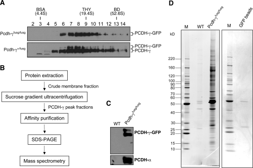
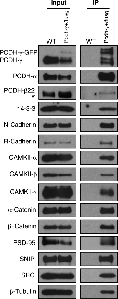
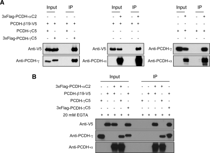
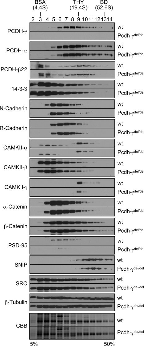
The three tandem-arrayed protocadherin (Pcdh) gene clusters, namely Pcdh-alpha, Pcdh-beta, and Pcdh-gamma, play important roles in the development of the vertebrate central nervous system. To gain insight into the molecular action of PCDHs, we performed a systematic proteomics analysis of PCDH-gamma-associated protein complexes. We identified a list of 154 non-redundant proteins in the PCDH-gamma complexes. This list includes nearly 30 members of clustered Pcdh-alpha, -beta, and -gamma families as core components of the complexes and additionally over 120 putative PCDH-associated proteins. We validated a selected subset of PCDH-gamma-associated proteins using specific antibodies. Analysis of the identities of PCDH-associated proteins showed that the majority of them overlap with the proteomic profile of postsynaptic density preparations. Further analysis of membrane protein complexes revealed that several validated PCDH-gamma-associated proteins exhibit reduced levels in Pcdh-gamma-deficient brain tissues. Therefore, PCDH-gamma s are required for the integrity of the complexes. However, the size of the overall complexes and the abundance of many other proteins remained unchanged, raising a possibility that PCDH-alphas and PCDH-betas might compensate for PCDH-gamma function in complex formation. As a test of this idea, RNA interference knockdown of both PCDH-alphas and PCDH-gamma s showed that PCDHs have redundant functions in regulating neuronal survival in the chicken spinal cord. Taken together, our data provide evidence that clustered PCDHs coexist in large protein complexes and have overlapping functions during vertebrate neural development.


Figures

Fig. 1.
Fig. 1.
Analysis of PCDH-γ macromolecular protein complexes using sucrose gradient ultracentrifugation and 2D BN/SDS-PAGE. A, SG ultracentrifugation analysis of PCDH-γ complexes. Brain membrane protein extracts from P0 or P21 mice were subjected to ultracentrifugation on a 5–50% sucrose gradient. Wild-type (WT; P0) membrane proteins were also treated with 1% SDS before SG ultracentrifugation to dissociate non-covalently linked PCDH-γ complexes. Samples from Pcdh-γdel/del mice served as a negative control to show the specificity of Western blot analysis. B, 2D BN/SDS-PAGE analysis of PCDH-γ complexes. Crude membrane protein extracts were subjected to 2D BN/SDS-PAGE analysis followed by Western blot analysis using an anti-pan-PCDH-γ antibody. The NativeMark unstained protein standard (Invitrogen) was used as molecular mass standard. The arrowhead in B indicates protein aggregation at the gel entry point (well bottom). THY, thyroglobulin; BD, blue dextran.
Fig. 2.
Fig. 2.
Affinity purification of PCDH-γ protein complexes. A, SG analysis of PCDH-γ-GFPs complexes from Pcdh-γ-GFPfusg/fusg and Pcdh-γ-GFP+/fusg mice (P0). PCDH-γ-GFPs and wild-type PCDH-γs are indicated. B, the flow chart of purification procedures. C, Western blot analysis of PCDH-γ-GFP immune complexes purified from Pcdh-γ-GFPfusg/fusg brains (P0–P5). An equal number of wild-type (WT) mouse brains served as a negative control for the purification. D, SYPRO Ruby-stained SDS-PAGE of the purified PCDH-γ-GFP immune complexes (left). To examine the possible protein contamination from anti-GFP-agarose beads, an equal amount of empty beads was eluted followed by SDS-PAGE and SYPRO Ruby staining (right). Only two faint bands, IgG heavy and light chains, were detected. M indicates the lanes loaded with marker proteins. THY, thyroglobulin; BD, blue dextran.
Fig. 3.
Fig. 3.
Confirmation of mass spectrometry-identified proteins using Western blots. The crude membrane protein extracts from P21 Pcdh-γ+/fusg mouse brains were immunoprecipitated with anti-GFP beads. The purified immune complexes were used for Western blot analyses using antibodies against PCDH-γs, PCDH-αs, PCDH-β22, 14-3-3, N-cadherin, R-cadherin, CAMKII-α, CAMKII-β, CAMKII-γ, α-catenin, β-catenin, PSD-95, SNIP, SRC family kinases, and β-tubulin. Wild-type (WT) mouse brains were used as a negative control. Inputs are equal amounts of the crude membrane protein extracts from both genotypes. “*” marks a nonspecific band partially overlapped with PCDH-β22 (also seen in Fig. 5).
Fig. 4.
Fig. 4.
PCDH-α, PCDH-β, and PCDH-γ isoforms interact with each other in vitro. A, co-IP experiments show that PCDH-α isoform C2, PCDH-γ isoform C5, and PCDH-β isoform 22 interact with each other in transfected cells. HEK293T cells were co-transfected with different combinations of expression vectors followed by co-IP using the anti-FLAG M2 affinity resin and Western blot with anti-V5, anti-pan-PCDH-α, and anti-pan-PCDH-γ antibodies. B, interactions among different PCDH isoforms are calcium-independent. Similar experiments as in A were carried out with an addition of 20 mm EGTA in the lysis buffer to deplete calcium.
Fig. 5.
Fig. 5.
Comparison of PCDH-α, PCDH-β, and PCDH-γ protein complexes from mouse brain using SG and 2D BN/SDS-PAGE. The SG and 2D BN/SDS-PAGE profiles of PCDH-γs, PCDH-αs, PCDH-β22, and other marker proteins are compared using P0 mouse brain samples. The SG and 2D BN/SDS-PAGE analyses were performed as in Fig. 1 with the indicated antibodies. “*” indicates nonspecific bands detected by anti-PCDH-α or anti-PCDH-β22 antibody. A, SG analysis. B, 2D BN/SDS-PAGE analysis. THY, thyroglobulin; BD, blue dextran; N-CAM, neural cell adhesion molecule.
Fig. 6.
Fig. 6.
Sucrose gradient analyses of mass spectrometry-identified proteins in wild-type and Pcdh-γdel/del mice. Equal amounts of membrane protein extracts from neonatal wild-type (wt) and Pcdh-γdel/del mouse brains were analyzed on a 5–50% sucrose gradient by ultracentrifugation. Equal amounts of individual fractions were separated by SDS-PAGE and analyzed by Western blots using antibodies against PCDH-γs, PCDH-αs, PCDH-β22, 14-3-3, N-cadherin, R-cadherin, CAMKII-α, CAMKII-β, CAMKII-γ, α-catenin, β-catenin, PSD-95, SNIP, SRC family kinases, and β-tubulin. The Coomassie Brilliant Blue (CBB)-stained gels of both wild-type and Pcdh-γdel/del at the area of around 55 kDa were used as loading controls to show an equal amount of proteins loaded. Each pair of Western blots from two genotypes were probed and developed at the same time. THY, thyroglobulin; BD, blue dextran.
Fig. 7.
Fig. 7.
PCDH-γs and PCDH-αs have overlapping function in regulating neuronal survival in developing chicken spinal cord. A, a diagram showing the PB transposon expressing shRNAs and the Ff-Luc reporter to test shRNA knockdown. B, relative efficiency of shRNA knockdown against the chicken PCDH-γ (left) or PCDH-α target sequence (right). HeLa cells were co-transfected with the Ff-Luc PCDH-α or PCDH-γ target reporter, individual effector shRNAs against PCDH-α or PCDH-γ, and Renilla luciferase (Rn-Luc). The relative knockdown efficiency is reflected by relative ratios of Ff/Rn-Luc activity using a GFP shRNA as the negative control. Shown are triplicates of transfection experiments. Bars show standard errors of the mean (SEM). The more effective shRNAs (γ-si1, γ-si3, α-si1, and α-si22) were identified for subsequent studies. C, to knock down endogenous chicken PCDH-αs and PCDH-γs, individual PB shRNAs were co-electroporated with a PBase expression vector into chicken neural tube at stage 12. The spinal cord was dissected into the electroporated side (E) and non-electroporated side (C) 4 days after electroporation (EP). The dissected tissues were used for detecting PCDH-αs and PCDH-γs. Both PCDH proteins are reduced in the electroporated side of the spinal cord. Note that residual levels of PCDH proteins remain possibly due to incompleteness of both electroporation and RNAi knockdown. D and E, simultaneous knockdown of PCDH-α and PCDH-γ induces neuronal apoptosis in the spinal cord. Different combinations of the indicated shRNAs were electroporated into one side of the spinal cord. Cryostat sections of the electroporated spinal cords were double stained with anti-active caspase-3 (red) and anti-GFP (green). D, representative images of active caspase-3-positive cells in the PCDH shRNA electroporated spinal cord (left in each panel). The yellow arrowheads mark the apoptotic cells. Composite images double stained with anti-GFP (green) and 4′,6-diamidino-2-phenylindole (blue) are shown for each panel on the right, demonstrating the stable transposition efficiency. E, quantitative analysis of cell death in the shRNA-expressing spinal cord. To exclude some apoptotic cells (mainly motoneurons) during normal development, Cell death index = Number of caspase-3-positive cells per section on the electroporated side − Number of caspase-3-positive cells per section on the non-electroporated side. Plotted are average cell death index numbers from individual electroporated chicken embryos. For each embryo, five randomly picked GFP-positive sections were used to generate the average cell death index. The results were subjected to a two-tail t test, and p values <0.0001 were obtained between the three combinations of PCDH-α and PCDH-γ shRNAs and other control samples. TR, translated region.

References

    1. Benson D. L., Colman D. R., Huntley G. W. (2001) Molecules, maps and synapse specificity. Nat. Rev. Neurosci. 2, 899–909 - PubMed
    1. Takeichi M. (2007) The cadherin superfamily in neuronal connections and interactions. Nat. Rev. Neurosci. 8, 11–20 - PubMed
    1. Sano K., Tanihara H., Heimark R. L, Obata S., Davidson M., St John T., Taketani S., Suzuki S. (1993) Protocadherins: a large family of cadherin-related molecules in central nervous system. EMBO J. 12, 2249–2256 - PMC - PubMed
    1. Morishita H., Yagi T. (2007) Protocadherin family: diversity, structure, and function. Curr. Opin. Cell Biol. 19, 584–592 - PubMed
    1. Halbleib J. M., Nelson W. J. (2006) Cadherins in development: cell adhesion, sorting, and tissue morphogenesis. Genes Dev. 20, 3199–3214 - PubMed

Publication types

MeSH terms