. 2009 Nov 1;15(21):6511-8.
doi: 10.1158/1078-0432.CCR-09-0565. Epub 2009 Oct 20.

Circulating mesothelin protein and cellular antimesothelin immunity in patients with pancreatic cancer

Affiliations

Circulating mesothelin protein and cellular antimesothelin immunity in patients with pancreatic cancer

Fabian Mc Johnston et al. Clin Cancer Res. .

Abstract

Purpose: Mesothelin is a glycoprotein expressed on normal mesothelial cells and is overexpressed in several histologic types of tumors including pancreatic adenocarcinomas. A soluble form of mesothelin has been detected in patients with ovarian cancer and malignant mesothelioma, and has prognostic value. Mesothelin has also been considered as a target for immune-based therapies. We conducted a study on the potential clinical utility of mesothelin as a biomarker for pancreatic disease and therapeutic target pancreatic cancer.

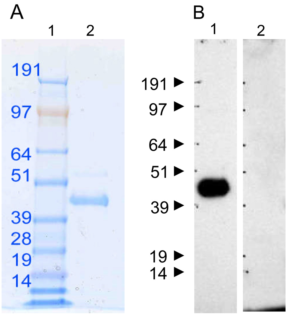
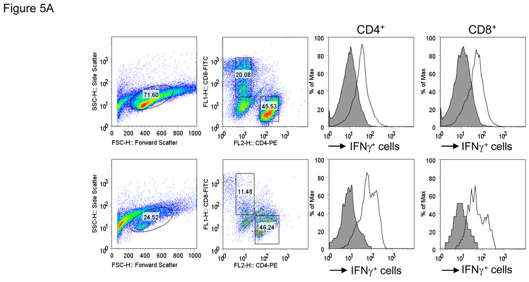
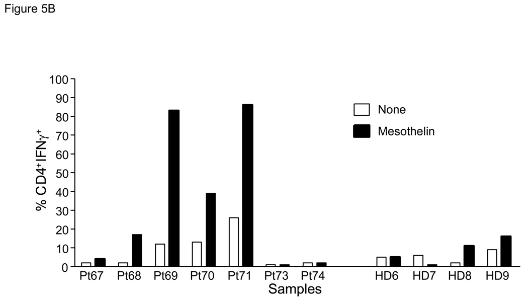
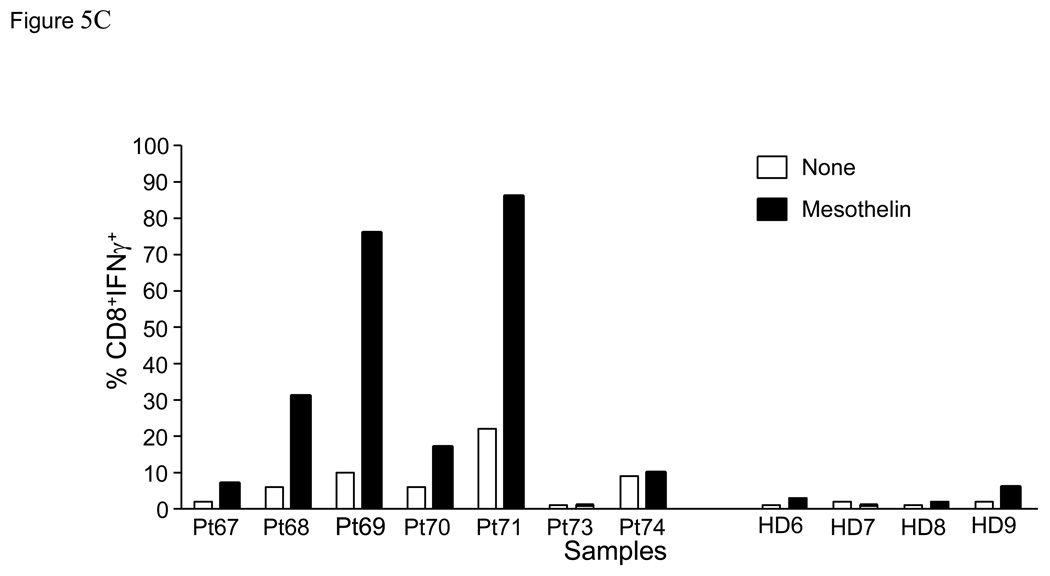
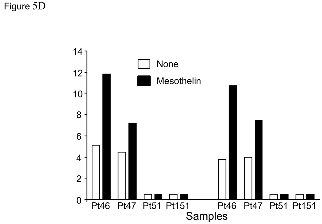
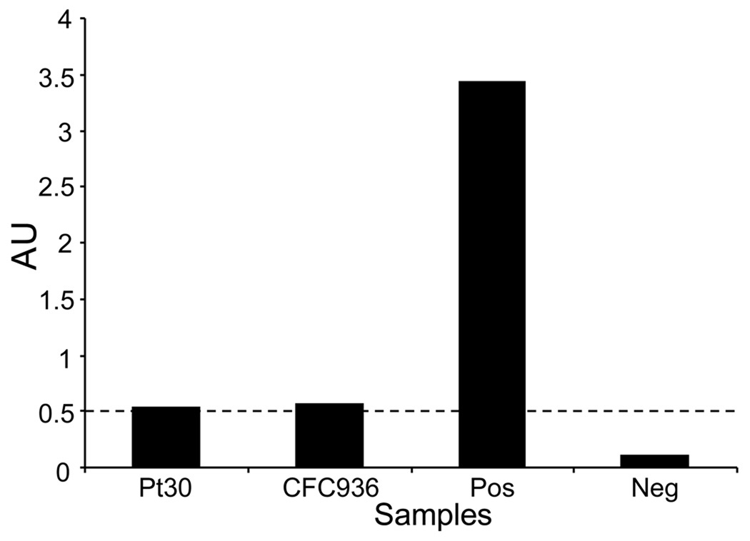
Experimental design: Tumor cell-bound and soluble mesothelin in patients was evaluated by immunohistochemistry and ELISA, respectively. The in vitro cellular immune response to mesothelin was evaluated by INF gamma ELISA and intracellular cytokine staining for IFN gamma in CD4(+) and CD8(+) T cells. The level of circulating antibodies to mesothelin was measured by ELISA.

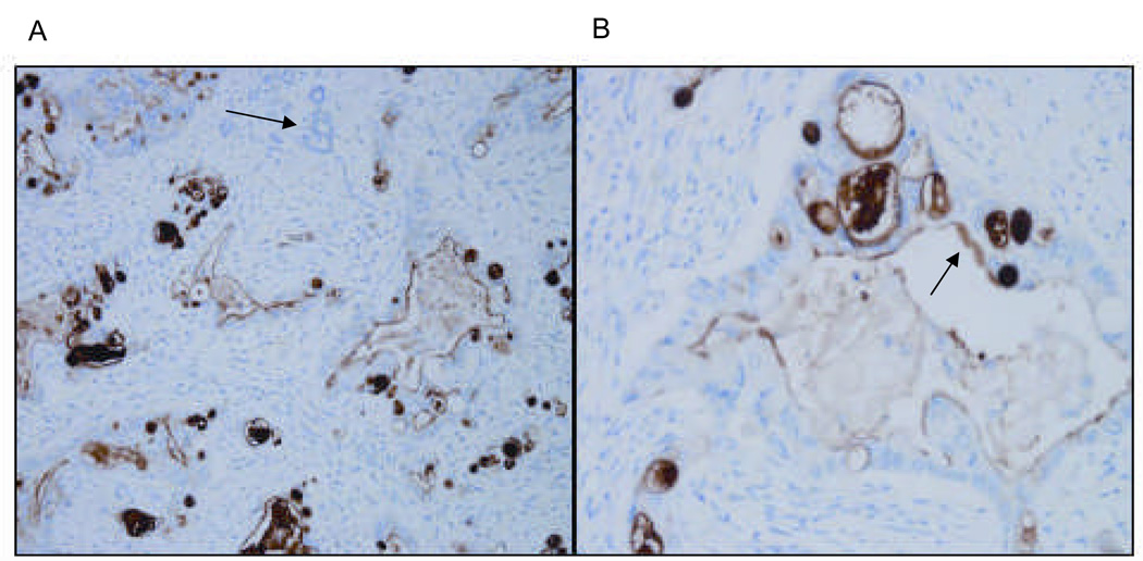
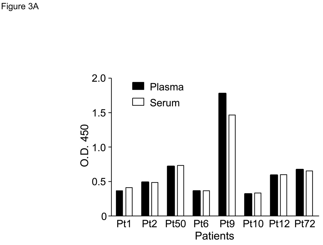
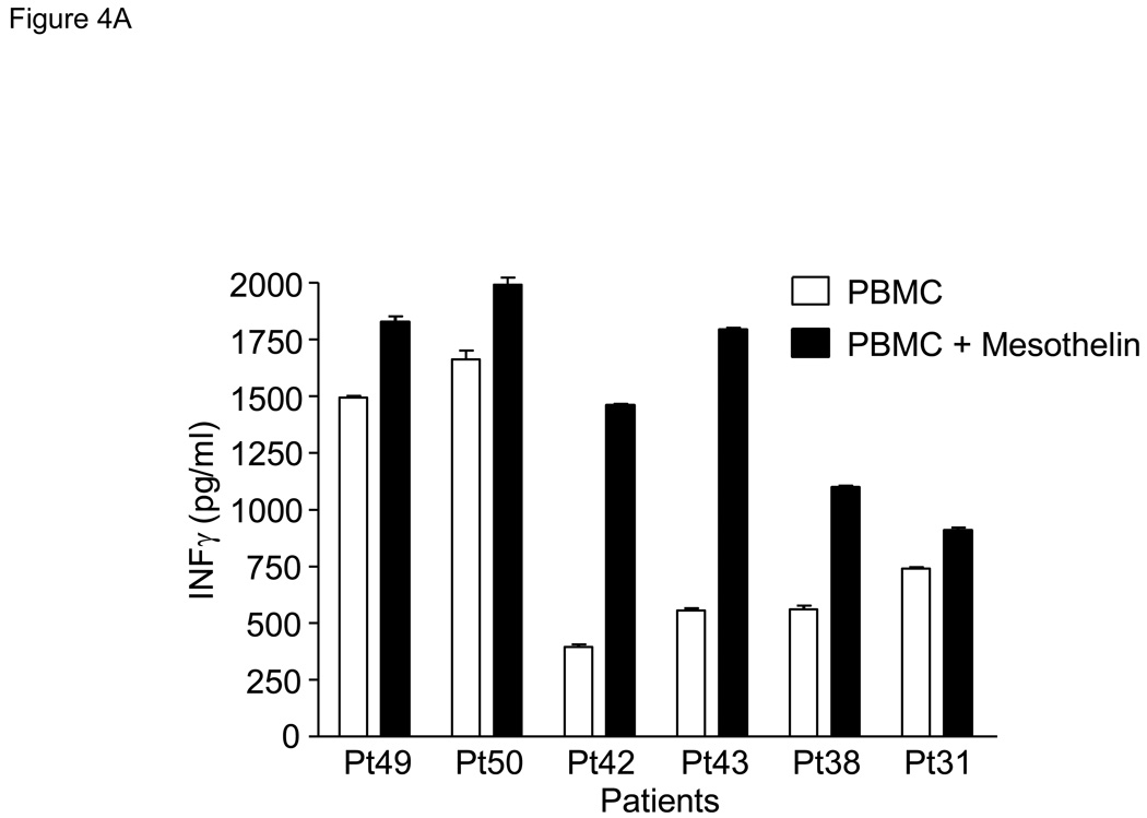
Results: All tumor tissue from patients with pancreatic adenocarcinoma expressed mesothelin (n = 10). Circulating mesothelin protein was detected in patients with pancreatic adenocarcinoma (73 of 74 patients) and benign pancreatic disease (5 of 5) but not in healthy individuals. Mesothelin-specific CD4(+) and CD8(+) T cells were generated from peripheral blood lymphocytes of patients with pancreatic cancer in 50% of patients compared with only 20% of healthy individuals. Antibodies reactive to mesothelin were detected in <3% of either patients or healthy individuals.

Conclusions: Circulating mesothelin is a useful biomarker for pancreatic disease. Furthermore, mesothelin-specific T cells can be induced in patients with pancreatic cancer. This suggests that mesothelin is a potential target for immune-based intervention strategies in pancreatic cancer.


Figures

Figure 1
Figure 1. Analysis of purified native mesothelin by gel electrophoresis and Western blot
A. Soluble mesothelin purified from culture supernatant of a mesothelioma cell line or urine from ovarian and pancreatic cancer patients(19) was run on a 4–12% SDS gel (lane 2) and stained by SYPRO-Ruby. Lane 1 was loaded with molecular weight markers. B. Two duplicate lanes were used for further development by Western blot using the anti-human mesothelin mAb, K1 in lane 1 or serum from a patient with pancreatic adenocarcinoma negative for anti-mesothelin antibody (lane 2).
Figure 2
Figure 2. Human pancreatic adenocarcinoma expresses mesothelin
Representative micrographs at low (A) and high (B) power of human pancreatic adenocarcinoma stained for mesothelin. Tissue sections were stained with the anti-human mesothelin mAb, 5B2 and counterstained by H&E. Non-tumor pancreatic glands are nonreactive with the mesothelin antibody, as shown by the arrow in panel A. Only tumor glands (see arrow in B) stain positive for mesothelin. The malignant tumor epithelium is 3+/3 positive in all the tumor glands.
Figure 3
Figure 3. Soluble mesothelin is detectable in serum and plasma of patients with pancreatic cancer
Data as determined by ELISA are expressed as absorption units (AU) at 450 nm using a 1:40 dilution. A. Comparison of serum and plasma for soluble mesothelin using samples from patients with metastatic pancreatic cancer. B. Circulating mesothelin levels in healthy volunteers, patients with benign pancreatic disease, patients with primary pancreatic cancer, and patients with unresectable pancreatic cancer. Each symbol represents a patient sample. Median values per group are indicated by a horizontal line. All median values in the patient groups are significantly elevated in comparison to that of healthy volunteers (p<0.05). The horizontal dashed line indicates the upper limit of negative values at AU = 0.2.
Figure 4
Figure 4. Patient-derived PBMC stimulated with mesothelin protein produce IFNγ
A. PBMC were obtained from 10 patients with pancreatic adenocarcinoma including poorly differentiated (Pts 16, 49 and 50), moderately differentiated (Pts18, 43 and 31), moderately to poorly differentiated (Pt42), and well-differentiated (Pts17, 38 and 40). PBMC were stimulated with mesothelin protein and cultured for 5–7 days followed by analysis of culture medium for IFNγ by ELISA. Control PBMC were cultured in medium alone. As a group, the mesothelin cultures contained significantly more IFNγ than the unstimulated cultures (p<0.05). B. After a single stimulation with mesothelin protein, PBMC were taken out of culture, washed, and placed overnight medium without mesothelin. The next day PBMC were restimulated with irradiated PBMC or PBMC pulsed with mesothelin. Culture medium was tested for the presence of IFNγ by ELISA. The mesothelin stimulated cultures but not the mammaglobin-stimulated cultures contained significantly more IFNγ than the unstimulated cultures, p<0.05.
Figure 5
Figure 5. Mesothelin-specific T cells detected by intracellular IFNγ
PBMC were obtained from seven patients with pancreatic adenocarcinoma including three patients with moderately differentiated tumors (Pts67, 73 and 74), two with moderately to poorly differentiated tumor (Pts69 and 71), and two with poorly differentiated tumors (Pts68 and 70) A. Representative FACS analyses of patient derived mesothelin-specific CD8 and CD4 T cells. The percentage IFNγ+ lymphocytes was determined by gating on either CD4+ or CD8+ cells. Mesothelin-stimulated PBMC were rested overnight and restimulated with autologous PBMC (shaded) or PBMC pulsed with mesothelin protein (unshaded). The percentage IFNγ+ cells was subsequently determined in both cultures. B and C: data summary showing percent double positive cells for INFγ and T cell markers CD4 (B) and CD8 (C). 4/8 patients with pancreatic adenocarcinoma showed significantly elevated levels of IFNγ in both T cell populations. There is a minimal response in 2/4 healthy, age-matched donors in the CD4 cell population. D. Summary data using PBMC from four patients with benign pancreatic lesions, IPMN. Pts47, 51, and 151 contained benign lesions without invasion of tumor whereas the lesion from patient 46 was found to contain small loci of invasive cancer.
Figure 6
Figure 6. Detection of anti-mesothelin antibodies in serum of patients with pancreatic adenocarcinoma
Serum from 56 patients and 35 healthy controls was tested for the presence of anti-human mesothelin IgG antibodies by ELISA. Serum from only one patient and one healthy control tested positive, albeit weakly, defined as an AU ≥ 0.5 at a 1:20 diluted sample(19). BSA was added as a negative control, and validated serum from a patient with ovarian carcinoma(19) was included as a positive control.

References

    1. Jemal A, Siegel R, Ward E, Hao Y, Xu J, Murray T, et al. Cancer statistics, 2008. CA Cancer J.Clin. 2008;58:71–96. - PubMed
    1. Kubuschok B, Neumann F, Breit R, Sester M, Schormann C, Wagner C, et al. Naturally occurring T-cell response against mutated p21 ras oncoprotein in pancreatic cancer. Clin.Cancer Res. 2006;12:1365–1372. - PubMed
    1. Schmitz-Winnenthal FH, Volk C, Z'graggen K, Galindo L, Nummer D, Ziouta Y, et al. High frequencies of functional tumor-reactive T cells in bone marrow and blood of pancreatic cancer patients. Cancer Res. 2005;65:10079–10087. - PubMed
    1. Schmitz-Winnenthal FH, Escobedo LV, Beckhove P, Schirrmacher V, Bucur M, Ziouta Y, et al. Specific immune recognition of pancreatic carcinoma by patient-derived CD4 and CD8 T cells and its improvement by interferon-gamma. Int.J.Oncol. 2006;28:1419–1428. - PubMed
    1. Fukunaga A, Miyamoto M, Cho Y, Murakami S, Kawarada Y, Oshikiri T, et al. CD8+ tumor-infiltrating lymphocytes together with CD4+ tumor-infiltrating lymphocytes and dendritic cells improve the prognosis of patients with pancreatic adenocarcinoma. Pancreas. 2004;28:e26–e31. - PubMed

Publication types