

Dose to radiosensitive organs during routine chest CT: effects of tube current modulation
- PMID: 19843751
- PMCID: PMC2954276
- DOI: 10.2214/AJR.09.2886
Dose to radiosensitive organs during routine chest CT: effects of tube current modulation
Abstract
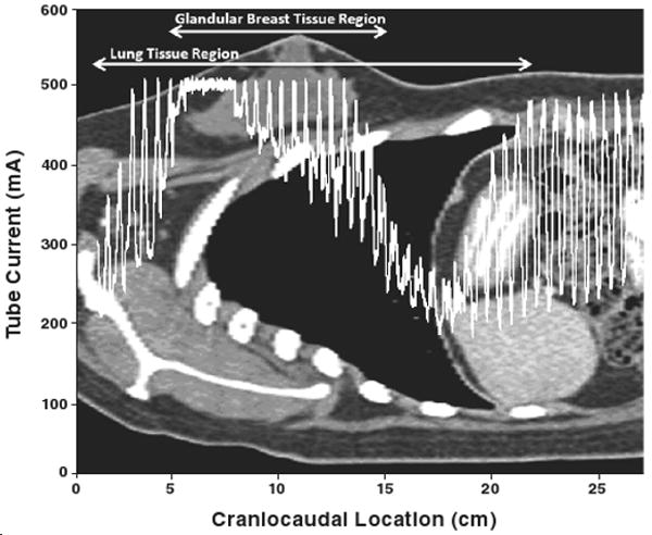
Objective: The aims of this study were to estimate the dose to radiosensitive organs (glandular breast and lung) in patients of various sizes undergoing routine chest CT examinations with and without tube current modulation; to quantify the effect of tube current modulation on organ dose; and to investigate the relation between patient size and organ dose to breast and lung resulting from chest CT examinations.
Materials and methods: Thirty voxelized models generated from images of patients were extended to include lung contours and were used to represent a cohort of women of various sizes. Monte Carlo simulation-based virtual MDCT scanners had been used in a previous study to estimate breast dose from simulations of a fixed-tube-current and a tube current-modulated chest CT examinations of each patient model. In this study, lung doses were estimated for each simulated examination, and the percentage organ dose reduction attributed to tube current modulation was correlated with patient size for both glandular breast and lung tissues.
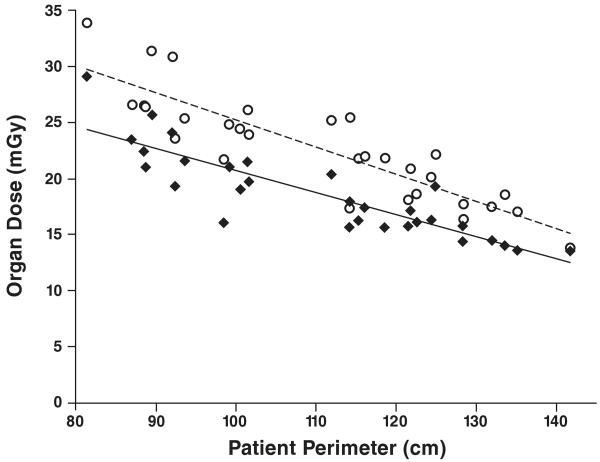
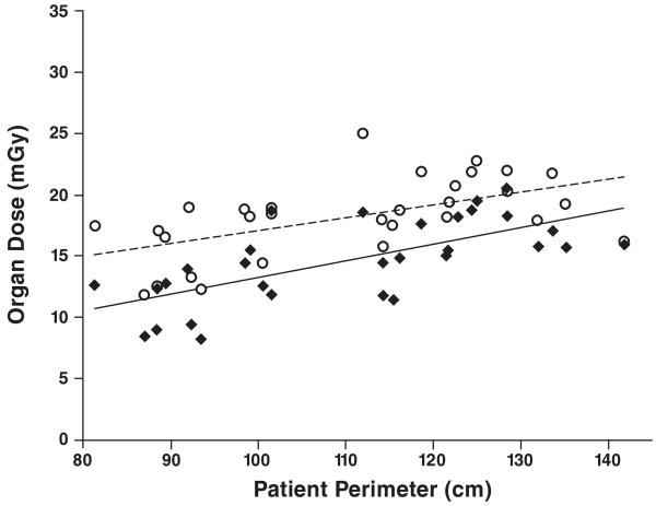
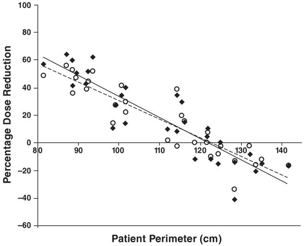
Results: The average radiation dose to lung tissue from a chest CT scan obtained with fixed tube current was 23 mGy. The use of tube current modulation reduced the lung dose an average of 16%. Reductions in organ dose (up to 56% for lung) due to tube current modulation were more substantial among smaller patients than larger. For some larger patients, use of tube current modulation for chest CT resulted in an increase in organ dose to the lung as high as 33%. For chest CT, lung dose and breast dose estimates had similar correlations with patient size. On average the two organs receive approximately the same dose effects from tube current modulation.
Conclusion: The dose to radiosensitive organs during fixed-tube-current and tube current-modulated chest CT can be estimated on the basis of patient size. Organ dose generally decreases with the use of tube current-modulated acquisition, but patient size can directly affect the dose reduction achieved.
Figures
References
-
- Mettler FA, Jr, Thomadsen BR, Bhargavan M, et al. Medical radiation exposure in the U.S. in 2006: preliminary results. Health Phys. 2008;95:502–507. - PubMed
-
- Hurwitz LM, Yoshizumi TT, Reiman RE, et al. Radiation dose to the female breast from 16-MDCT body protocols. AJR. 2006;186:1718–1722. - PubMed
-
- Mayo JR, Aldrich J, Muller NL. Radiation exposure at chest CT: a statement of the Fleischner Society. Radiology. 2003;228:15–21. - PubMed
-
- The 2007 recommendations of the International Commission on Radiological Protection: ICRP publication 103. Ann ICRP. 2007;37:1–332. No authors listed. - PubMed
-
- Preston DL, Ron E, Tokuoka S, et al. Solid cancer incidence in atomic bomb survivors: 1958–1998. Radiat Res. 2007;168:1–64. - PubMed
Publication types
MeSH terms
Grants and funding
LinkOut - more resources
Full Text Sources
Medical
