Comparative Study
. 2009 Nov 1;183(9):5615-21.
doi: 10.4049/jimmunol.0901691.

Antibodies to the desmoglein 1 precursor proprotein but not to the mature cell surface protein cloned from individuals without pemphigus

Affiliations
Comparative Study

Antibodies to the desmoglein 1 precursor proprotein but not to the mature cell surface protein cloned from individuals without pemphigus

Jun Yamagami et al. J Immunol. .

Abstract

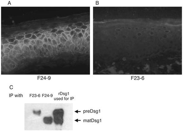
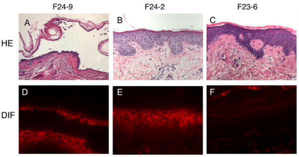
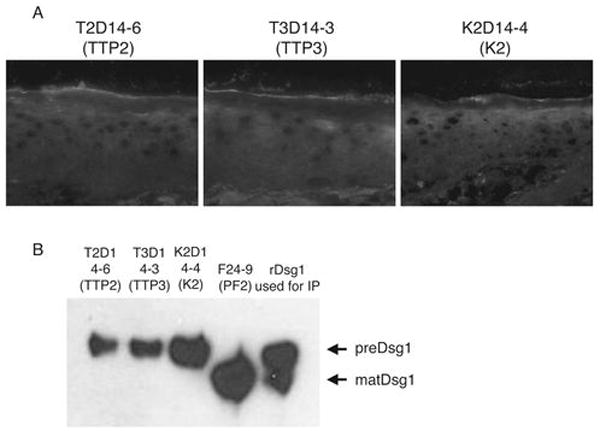
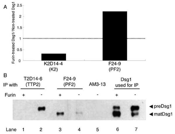
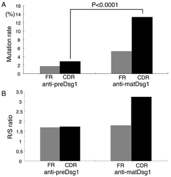
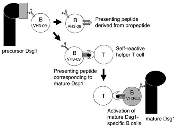
In pemphigus foliaceus (PF), autoantibodies against desmoglein 1 (Dsg1) cause blisters. Using Ab phage display, we have cloned mAbs from a PF patient. These mAbs, like those from a previous patient, were directed against mature Dsg1 (matDsg1) on the cell surface of keratinocytes and precursor Dsg1 (preDsg1) in the cytoplasm. To determine whether individuals without pemphigus have B cell tolerance to Dsg1, we cloned mAbs from two patients with thrombotic thrombocytopenic purpura and a healthy person. We found mAbs against preDsg1, but not matDsg1. All but 1 of the 23 anti-preDsg1 mAbs from PF patients and those without PF used the VH3-09 (or closely related VH3-20) H chain gene, whereas no PF anti-matDsg1 used these genes. V(H) cDNA encoding anti-preDsg1 had significantly fewer somatic mutations than did anti-matDsg1 cDNA, consistent with chronic Ag-driven hypermutation of the latter compared with the former. These data indicate that individuals without PF do not have B cell tolerance to preDsg1 and that loss of tolerance to matDsg1 is not due to epitope shifting of anti-preDsg1 B cells (because of different V(H) gene usage). However, presentation of peptides from Dsg1 by preDsg1-specific B cells may be one step in developing autoimmunity in PF.


Conflict of interest statement

Disclosures: The authors declare no conflict of interest.

Figures

Figure 1
Figure 1
Immunohistological and immunochemical analysis of two major types of anti-Dsg1 mAbs isolated from PF patient 2 (PF2). A and B, IIF on normal human skin with anti-HA of F24-9 and F23-6 mAbs (each cloned with an HA tag). F24-9 stains the cell surface of keratinocytes whereas F23-6 does not stain the cell surface but, if it stains at all, weakly stains the cytoplasm, suggesting binding of mat and pre Dsg1, respectively. C, Immunoblotting with anti-E-tag. Left 2 lanes show immunoblot of an immunoprecipitation of recombinant human Dsg1Ehis. F23-6 precipitates a higher molecular weight band, preDsg1, while F24-9 binds to lower molecular weight matDsg1. Right lane shows immunoblot of recombinant Dsg1Ehis used for the immunoprecipitation and shows both the high and lower molecular weight species.
Figure 2
Figure 2
Pathogenicity and binding of mAbs from patient PF2 as determined by injection into normal human skin in organ culture. A and D, F24-9 causes a blister in the superficial epidermis typical of PF (as shown by histology with routine hematoxylin and eosin staining) and shows typical keratinocyte cell surface staining by direct immunofluorescence (DIF) of the injected skin. B and E, F24-2 does not cause an epidermal blister but binds the cell surface of keratinocytes. C and F, F23-6 is non-pathogenic and shows no binding by DIF. These results demonstrate the three types of antibodies cloned from patient PF2: pathogenic anti-matDsg1 (F24-9), non-pathogenic anti-matDsg1 (F24-2), and non-pathogenic anti-preDsg1 (F23-6).
Figure 3
Figure 3
IIF and immunoprecipitation of mAbs cloned from TTP patients (TTP2, TTP3) and a normal individual (K2). A, All of the anti-Dsg1 mAbs isolated from non-pemphigus individuals show either no staining or very weak cytoplasmic staining in IIF of normal human skin. None show cell surface staining. B, These mAbs immunoprecipitate recombinant preDsg1, in contrast to F24-9, a mAb cloned from patient PF2, that binds the keratinocyte cell surface (Fig. 1A) and immunoprecipitates matDsg1.
Figure 4
Figure 4
Furin, which processes preDsg1 to matDsg1, causes decreased binding of anti-Dsg1 antibodies cloned from individuals without pemphigus. A, Furin treatment of Dsg1 ELISA plates causes decreased binding of K2D14-4, a mAb from a normal individual, but increased binding of F24-9, a pathogenic cell surface mAb from patient PF2. B, Immunoblot with anti-E-tag. Dsg1Ehis recombinant protein without furin treatment shows a high and lower molecular weight band (lane 7). Treatment with furin shows decreased intensity of the higher molecular weight band from processing of preDsg1 to matDsg1 (lane 6). T2D14-6 immunoprecipitates preDsg1 (lane 2) that is not longer detectable after furin treatment (lane 1). However, F24-9 precipitates only matDsg1 with or without furin treatment (lanes 3,4).
Figure 5
Figure 5
Variable heavy chains of anti-matDsg1 mAbs have higher mutation rates compared to the VH germline sequences and a higher replacement to silent (R/S) ratio of those mutations than anti-preDsg1 mAbs. A, Comparison of the average mutation rates shows that anti-matDsg1 antibodies have more mutations than anti-preDsg1 antibodies throughout the heavy chain variable regions and that these mutations occur more often in CDRs than in FR regions. B, The replacement to silent ratio of somatic mutations is elevated in the CDRs compared to FRs of anti-matDsg1 antibodies.
Figure 6
Figure 6
A model of PF development in which, as a first step, anti-preDsg1 B cells present peptides from matDsg1. These B cells could present antigen to T cells that recognize matDsg1 peptides. Once such T cells proliferate they can then provide help, not only to the original anti-preDsg1 B cells but also to any B cells that have escaped from tolerance to matDsg1. Such a model requires both T cells and B cells to escape tolerance to matDsg1, unlikely events consistent with the rarity of PF. Note that preDsg1 reacting B cells and matDsg1-specific B cells have different clonal origins, consistent with our data showing that anti-preDsg1 and anti-matDsg1 cloned mAbs derive from different B cell clones.

References

    1. Stanley JR, Amagai M. Pemphigus, bullous impetigo, and staphylococcal scalded skin syndrome. N Engl J Med. 2006;355:1800–1810. - PubMed
    1. Eyre RW, Stanley JR. Human autoantibodies against a desmosomal protein complex with a calcium-sensitive epitope are characteristic of pemphigus foliaceus patients. J Exp Med. 1987;165:1719–1724. - PMC - PubMed
    1. Koulu L, Kusumi A, Steinberg MS, Klaus Kovtun V, Stanley JR. Human autoantibodies against a desmosomal core protein in pemphigus foliaceus. J Exp Med. 1984;160:1509–1518. - PMC - PubMed
    1. Posthaus H, Dubois CM, Muller E. Novel insights into cadherin processing by subtilisin-like convertases. FEBS Lett. 2003;536:203–208. - PubMed
    1. Wahl JK, III, Kim YJ, Cullen JM, Johnson KR, Wheelock MJ. N-cadherin-catenin complexes form prior to cleavage of the proregion and transport to the plasma membrane. J Biol Chem. 2003;278:17269–17276. - PubMed

Publication types

MeSH terms