. 2009 Oct 21;4(10):e7535.
doi: 10.1371/journal.pone.0007535.

MicroRNA miR-378 regulates nephronectin expression modulating osteoblast differentiation by targeting GalNT-7

Affiliations

MicroRNA miR-378 regulates nephronectin expression modulating osteoblast differentiation by targeting GalNT-7

Shireen Kahai et al. PLoS One. .

Abstract

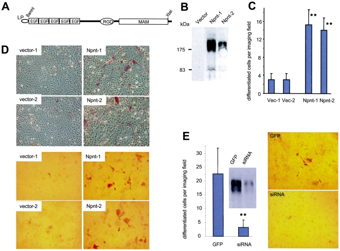
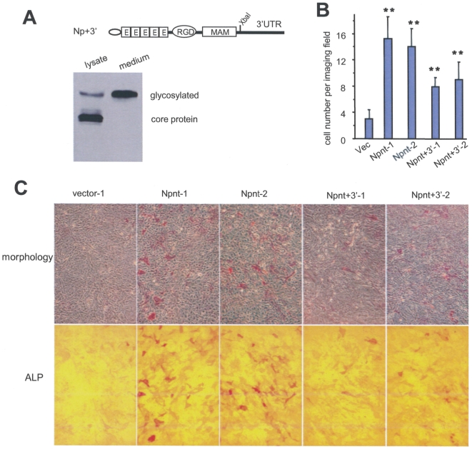
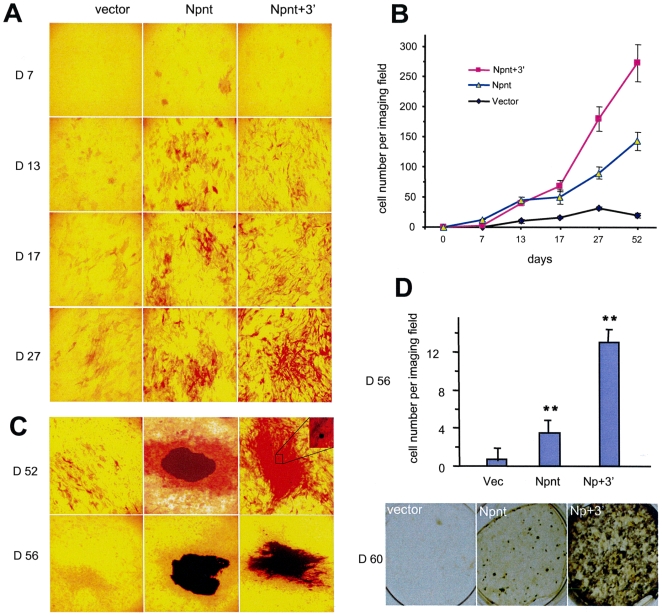
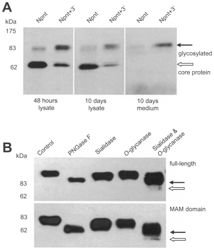
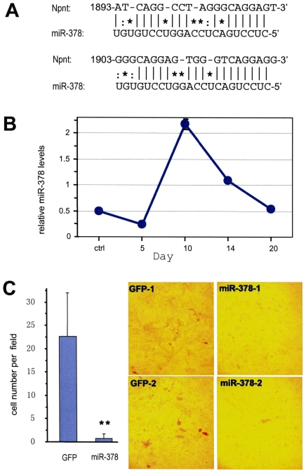
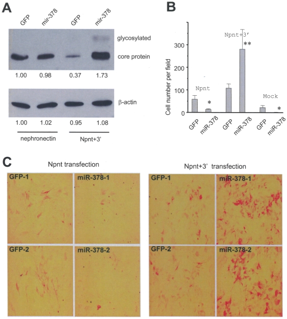
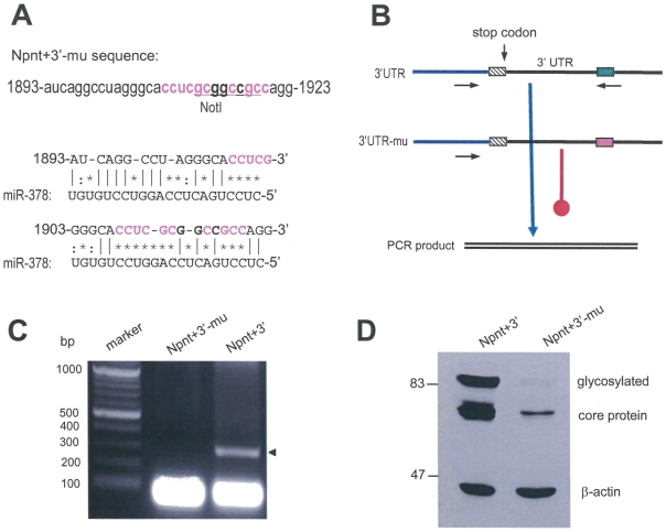
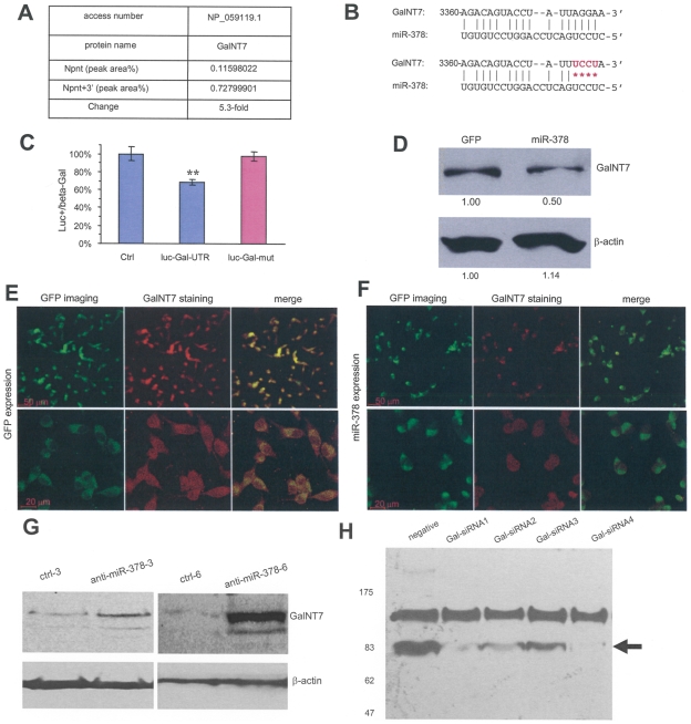
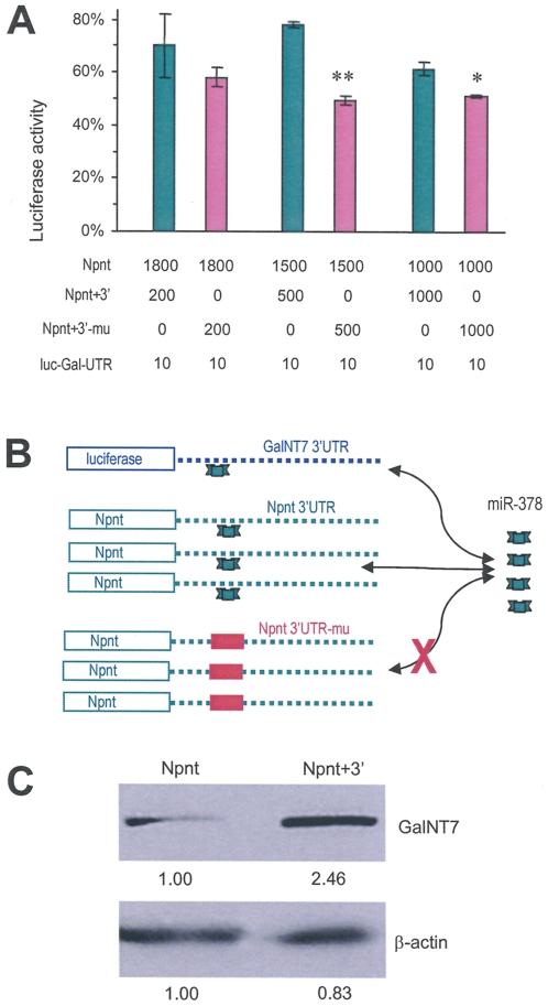
MicroRNAs (miRNAs) are small fragments of single-stranded RNA containing 18-24 nucleotides, and are generated from endogenous transcripts. MicroRNAs function in post-transcriptional gene silencing by targeting the 3'-untranslated region (UTR) of mRNAs, resulting in translational repression. We have developed a system to study the role of miRNAs in cell differentiation. We have found that one of the miRNAs tested in our system (miR-378, also called miR-378*) plays a role in modulating nephronectin-mediated differentiation in the osteoblastic cell line, MC3T3-E1. Nephronectin is an extracellular matrix protein, and we have demonstrated that its over-expression enhanced osteoblast differentiation and bone nodule formation. Furthermore, we found that the nephronectin 3'-untranslated region (3'UTR) contains a binding site for miR-378. Stable transfection of MC3T3-E1 cells with miR-378 inhibited cell differentiation. MC3T3-E1 cells stably transfected with nephronectin exhibited higher rates of differentiation and nodule formation as compared with cells transfected with nephronectin containing the 3'UTR in the early stages of development, suggesting that endogenous miR-378 is present and active. However, in the later stages of MC3T3-E1 development, the differentiation rates were opposite, with higher rates of differentiation and nodule formation in the cells over-expressing the 3'UTR of nephronectin. This appeared to be the consequence of competition between nephronectin and UDP-N-acetyl-alpha-D-galactosamine:polypeptide N-acetylgalactosaminyltransferase 7 (GalNAc-T7 or GalNT7) for miR-378 binding, resulting in increased GalNT7 activity, which in turn lead to increased nephronectin glycosylation and product secretion, thereby resulting in a higher rate of osteoblast differentiation.


Conflict of interest statement

Competing Interests: The authors have declared that no competing interests exist.

Figures

Figure 1
Figure 1. Alkaline phosphatase expression in MC3T3-E1 cells over-expressing nephronectin.
(A) A full-length nephronectin construct was generated containing the 5 EGF-like repeats, RGD domain, and the MAM domain. A leading peptide (LP) was tagged at the N-terminal region of the construct (Panel A). (B) After transfection, conditioned medium from two nephronectin- and one vector-transfected MC3T3-E1 clones was analyzed for stable expression and secretion of the full-length nephronectin protein by western blot, using the monoclonal antibody 4B6 which recognizes an epitope in the leading peptide. (C) Two nephronectin- and two vector-transfected MC3T3-E1 clones were grown for 10 days and stained for ALP expression. Multiple fields were examined for each construct, and stained cells were counted per field for a quantitative graphical representation. Over-expressing nephronectin (Npnt-1 and Npnt-2) enhanced ALP expression. (D) Typical micrographs of ALP expression in the two clones were taken using two different phases (top and bottom panels) of the microscope for better estimation of ALP activity. (E) Left, MC3T3-E1 cells stably transfected with an siRNA construct against nephronectin were grown in parallel with cells transfected with the empty vector for 10 days and then stained for ALP activity. Multiple fields were examined for each construct, and stained cells were counted per field for quantitative analysis. Inset shows the expression level of the nephronectin protein after transfection with the siRNA against nephronectin. Right, typical micrographs of ALP expression are shown.
Figure 2
Figure 2. The effect of nephronectin 3′UTR on osteoblast differentiation.
(A) A nephronectin construct with a fragment of the nephronectin 3′UTR spanning the target sequence of miR-378 is shown (Npnt+3′). Cell lysate and conditioned medium from MC3T3-E1 cells stably transfected with Npnt+3′ were analyzed by western blot showing secretion of the product. (B) Cells transfected with the full-length nephronectin (Npnt-1 and Npnt-2), nephronectin containing the 3′UTR (Npnt+3′-1 and Npnt+3′-2), or with the control vector were grown for 10 days and stained for ALP expression. Multiple fields were examined for each construct, and stained cells were counted per field for quantitative analysis. (C) Typical micrographs of ALP expression are shown using two different phases (top and bottom panels) of the microscope.
Figure 3
Figure 3. Nephronectin 3′UTR promotes nodule formation.
(A) Time course analysis showing the different effects of Npnt and Npnt+3′ transfection on MC3T3-E1 differentiation: early stages, Npnt>Npnt+3′; late stages, Npnt
Figure 4
Figure 4. The 3′UTR of nephronectin promoted nephronectin protein glycosylation.
(A) Cell lysate from the two MC3T3-E1 stable cells lines (over-expressing the nephronectin protein with and without the 3′UTR) was prepared from 48 hour cultures and 10 day cultures. The lysate and conditioned medium was analyzed on western blot probed with the 4B6 antibody. The bands representing glycosylated nephronectin and the nephronectin core protein have been marked separately. (B) Conditioned medium was treated with different de-glycosylation enzymes to reduce the observed molecular size in western blot to confirm nephronectin glycosylation. The cells transfected with either the full-length nephronectin or just the MAM domain possessed potential O-glycosylation, which accounts for the significant size-difference in Fig. 4A. Filled arrows indicate the de-O-glycosylated product; and open arrows show the size of core protein.
Figure 5
Figure 5. Targeting of nephronectin with miR-378.
(A) Computational algorithms were used to predict the miRNA that binds to a target sequence in the 3′-UTR of nephronectin. miR-378 was found to have the strongest binding to the 3′UTR of nephronectin from nucleotides 1902–1923. (B) Expression of endogenous miR-378 in MC3T3-E1 cells under differentiation-inducing conditions was measured by real-time PCR. (C) Left, MC3T3-E1 cells stably transfected with the miR-378 construct were grown in parallel with cells transfected with the control vector for 10 days and stained for ALP activity. Multiple fields were examined for each construct, and stained cells were counted per field for quantitative analysis. Right, typical micrographs of ALP expression are shown in two separate stable clones.
Figure 6
Figure 6. Effect of miR-378 and 3′UTR on nephronectin expression, and its biological activity in double-transfected cells.
(A) Western blot for Npnt expression in the double-transfected cells. miR-378 elevated Npnt expression only when 3′UTR was present. Both glycosylated and core protein bands were observed. (B) Cell differentiation as revealed by ALP staining for Npnt-transfected and Npnt+3′-transfected cells. miR-378 expression inhibited differentiation in Npnt- and mock-transfected cells. However, miR-378 promoted osteoblast differentiation extensively in Npnt+3′-transfected cells, implying that miR-378 interacted with the 3′UTR and exerted its effect. (C) Typical results in Panel B are provided showing that the presence of the 3′UTR promoted cell differentiation upon miR-378 transfection.
Figure 7
Figure 7. Mutation of the miR-378 binding sites inhibits nephronectin glycosylation.
(A) Mutation was generated in the 3′UTR of Npnt+3′ construct as shown (Npnt+3′-mu, upper panel). This impaired both putative binding sites for miR-378 (bottom). (B) Strategy of PCR showing interaction of miR-378 with Npnt 3′UTR, which was abolished by sited-directed mutagenesis. (C) Typical PCR products are shown using the mature miR-378 as a primer for the PCR (arrow). The lower bands were primers used for the PCR assays. (D) Cell lysate prepared from cells transfected with Npnt+3′ and the mutant Npnt+3′-mu was analyzed with western blot showing decreased expression of the mutant construct.
Figure 8
Figure 8. Targeting of O-glycosyltransferase, UDP-N-acetyl-alpha-D-galactosamine:polypeptide N-acetylgalactosaminyltransferase 7 (GalNT7) by miR-378.
(A) U87 cells transfected with Npnt or Npnt+3′ were harvested and subjected to proteomic analysis. Up-regulation of GalNT7 expression was seen in cells transfected with Npnt+3′. (B) Computational algorithms were used to predict potential targeting of miR-378 on GalNT7. miR-378 was found to possesses a potential target site on GalNT7 in nucleotides 3360-3378. A fragment of the GalNT7 3′UTR harbouring the miR-378 target site was inserted into a luciferase reporter vector. The seed region for miR-378 binding was mutated in GalNT7 as shown (red). (C) Luciferase activity assays were performed by co-transfection of miR-378 with luciferase reporter constructs. Decreased luciferase activity was detected in the construct containing GalNT7 3′UTR, which was abolished when the miR-378 target site was mutated. (n = 4, ** p<0.01). (D) Western blot showing decreased expression of GalNT7 in miR-378-transfected cells compared with GFP-transfected cells. (E-F) U87 cells stably transfected with GFP (E) or miR-378 (F) were immunostained with anti-GalNT7 antibody, followed by confocal microscopic visualization. While both types of cells expressed GFP (green), miR-378-transfected cells expressed lower levels of GalNT7 than the GFP-transfected cells (red). As a result, the merged color of miR-378-transfected cells were less yellow. (G) MC3T3 cells were transfected with anti-miR-378 or a control vector at 3 or 6 µg plasmids. Cell lysates were prepared and analyzed on western blot probed with anti-GalNT7 antibody. Transfection with anti-miR-378 enhanced GalNT7 expression. (H) MC3T3 cells were co-transfected with nephronectin and one of the four siRNA oligos against GalNT7 or an oligo at random sequence serving as a negative control. Culture medium was analyzed on western blot probed with the monoclonal antibody 4B6 that recognizes nephronectin. Transfection with siRNAs against GalNT7 decreased the levels of secreted nephronectin.
Figure 9
Figure 9. Npnt 3′UTR competes with GalNT7 3′UTR for miR-378 binding.
(A) U343 cells were transiently transfected with luciferase reporter vector harboring the GalNT7 3′UTR (lu-Gal-UTR) and different constructs at the amounts indicated on the figure (ng). Luciferase activities were normalized using the control as 100%. The luciferase activities were higher when co-transfected with Npnt+3′ as compared with Npnt+3′-mu, suggesting that increased supplies of Npnt+3′ absorbed some endogenous miRNAs freeing luciferase translation. (B) Diagram showing competition of Npnt+3′, but not Npnt+3′-mu, with GalNT7 for miR-378 binding. As a result, luc-Gal-UTR was freed from miR-378 binding and thereby having higher luciferase activities. (C) Western blot showing increased expression of GalNT7 in the Npnt+3′-transfected cells compared with the Npnt-transfected cells.

References

    1. Rubartelli A, Poggi A, Zocchi MR. The selective engulfment of apoptotic bodies by dendritic cells is mediated by the alpha(v)beta3 integrin and requires intracellular and extracellular calcium. Eur J Immunol. 1997;27:1893–1900. - PubMed
    1. Chendrimada TP, Gregory RI, Kumaraswamy E, Norman J, Cooch N, et al. TRBP recruits the Dicer complex to Ago2 for microRNA processing and gene silencing. Nature. 2005;436:740–744. - PMC - PubMed
    1. Hutvagner G, Zamore PD. A microRNA in a multiple-turnover RNAi enzyme complex. Science. 2002;297:2056–2060. - PubMed
    1. Lim LP, Glasner ME, Yekta S, Burge CB, Bartel DP. Vertebrate microRNA genes. Science. 2003;299:1540. - PubMed
    1. Lund E, Guttinger S, Calado A, Dahlberg JE, Kutay U. Nuclear export of microRNA precursors. Science. 2004;303:95–98. - PubMed

Publication types

LinkOut - more resources