. 2008 Jan 1:2:52-58.
doi: 10.2174/1874279300802010052.

Morphogenesis of Coronavirus HCoV-NL63 in Cell Culture: A Transmission Electron Microscopic Study

Affiliations

Morphogenesis of Coronavirus HCoV-NL63 in Cell Culture: A Transmission Electron Microscopic Study

Jan M Orenstein et al. Open Infect Dis J. .

Abstract

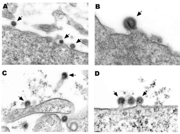
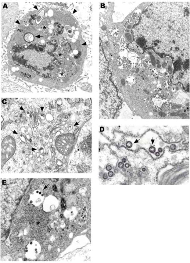
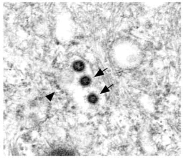
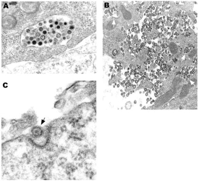
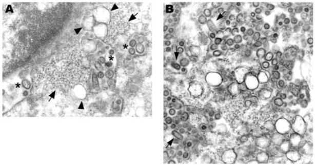
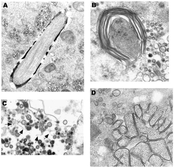
NL63 (HCoV-NL63) is a recently discovered human coronavirus that causes respiratory disease in infants and young children. NL63 productively infects LLCMK2 cells and ciliated epithelial cells of human airway cell cultures. Transmission electron microscopic (TEM) studies of NL63 infected LLCMK2 cells revealed that virions are spherical, spiked, and range from 75 to 115 nm in diameter. Virus replication predominantly occurs on the rough endoplasmic reticulum (RER), both perinuclear and cytoplasmic, and the Golgi. Plasma membrane budding was occasionally observed. As virus production increased, aberrant viral forms appeared with greater frequency. Unusual inclusions were present in infected cells including tubular and laminated structures. Pleomorphic double membrane-bound vesicles (DMV), measuring roughly 140 to 210 nm in diameter, were observed. The virus was released via exocytosis and cell lysis. In summary, we report the key morphologic characteristics of NL63 infection observed by TEM analysis.


Figures

Fig. 1
Fig. 1
(A) Three NL63 particles budding from the plasma membrane (arrows). (B) A single NL63 particle budding from the plasmalemma (arrow). (C) Two MHV particles budding from plasma membrane processes (arrows). (D) Two MHV particles budding from the plasmalemma (arrows) flank a particle that has completed budding. A × 76,000, B × 99,000, C × 79,000, D × 79,000.
Fig. 2
Fig. 2
(A) An electron dense, apoptotic cell rich in virus concentrate around the nucleus (stars). Two laminate bodies are present (arrows). There are several collections of granular nucleocapsid material (e.g., arrowheads). (B) Cytoplasmic and perinuclear vacuoles contain abundant virus. (C) Virions budding into Golgi cisternae (arrows). (D) Ribosomes are in place on a dilated RER cisternae containing virus. Electron dense granular nucleocapsid material can be seen in some of the virions (arrows). (E) Virions are budding into smooth membrane vacuoles. A × 8,500, B × 11,000, C × 30,000, D × 59,000, E × 25,000.
Fig. 3
Fig. 3
Three dense virions are seen in this RER cisternae still partially covered with ribosomes (arrowhead). Two of the virions have detectable spikes (arrows). × 64,000.
Fig. 4
Fig. 4
(A) Virions of varying density appear to be in the process of exocytosis from a healthy cell. (B) Virions are in vacuoles (arrows) near the plasma membrane and free trapped within complicated cell processes. The cell appears healthy, suggesting that the free virus was released by exocytosis. Some of the free virions are bullet-shaped. (C) A viral particle appears to be in the process of endocytosis in a coated pit. A × 50,000, B × 29,000, C × 120,000.
Fig. 5
Fig. 5
(A) This perinuclear field shows nucleocapsid material (arrow), vacuoles containing virus (stars), some bullet-shaped, and double-membrane vesicles (arrowheads). (B) Numerous double-membrane vesicles are surrounded by abundant viral particles, some bullet-shaped (arrows). A × 45,000, B × 48,000.
Fig. 6
Fig. 6
(A) This unusual inclusions contains longitudinally oriented tubules. Vacuoles containing virus are nearby. (B) Periodicity can be seen in a laminated structure that is in close proximity to pleomorphic viral particles (right side). (C) In the background are lightly staining tubular structures (arrows). Note the variability in virus diameter. (D) Granular nucleocapsid material is associated with electron dense structures that resemble linear arrays of “budding” virus. A x36,000, B x50,000, C x32,000, D x44,000.

References

    1. Ksiazek TG, Erdman D, Goldsmith CS, et al. A novel coronavirus associated with severe acute respiratory syndrome. N Engl J Med. 2003;348:1953–66. - PubMed
    1. Vicenzi E, Canducci F, Pinna D, et al. Coronaviridae and SARS-associated coronavirus strain HSR1. Emerg Infect Dis. 2004;10:413–8. - PMC - PubMed
    1. Goldsmith CS, Tatti KM, Ksiazek TG, et al. Ultrastructural characterization of SARS coronavirus. Emerg Infect Dis. 2004;10:320–6. - PMC - PubMed
    1. Sims AC, Baric RS, Yount B, Burkett SE, Collins PL, Pickles RJ. Severe acute respiratory syndrome coronavirus infection of human ciliated airway epithelia: role of ciliated cells in viral spread in the conducting airways of the lungs. J Virol. 2005;79:15511–24. - PMC - PubMed
    1. Peiris JSM, Lai ST, Poon LLM, et al. Coronavirus as a possible cause of severe acute respiratory syndrome. Lancet. 2003;361:1319–25. - PMC - PubMed

LinkOut - more resources