

Angiotensin AT1 receptor antagonism ameliorates murine retinal proteome changes induced by diabetes
- PMID: 19845401
- PMCID: PMC2798584
- DOI: 10.1021/pr9006415
Angiotensin AT1 receptor antagonism ameliorates murine retinal proteome changes induced by diabetes
Abstract
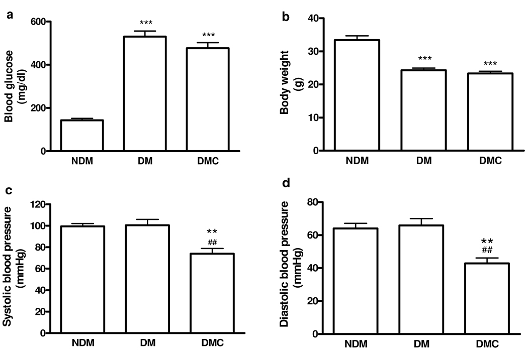
Diabetic retinopathy is the most common microvascular complication caused by diabetes mellitus and is a leading cause of vision loss among working-age adults in developed countries. Understanding the effects of diabetes on the retinal proteome may provide insights into factors and mechanisms responsible for this disease. We have performed a comprehensive proteomic analysis and comparison of retina from C57BL/6 mice with 2 months of streptozotocin-induced diabetes and age-matched nondiabetic control mice. To explore the role of the angiotensin AT1 receptor in the retinal proteome in diabetes, a subgroup of mice were treated with the AT1 antagonist candesartan. We identified 1792 proteins from retinal lysates, of which 65 proteins were differentially changed more than 2-fold in diabetic mice compared with nondiabetic mice. A majority (72%) of these protein changes were normalized by candesartan treatment. Most of the significantly changed proteins were associated with metabolism, oxidative phosphorylation, and apoptotic pathways. An analysis of the proteomics data revealed metabolic and apoptotic abnormalities in the retina from diabetic mice that were ameliorated with candesartan treatment. These results provide insight into the effects of diabetes on the retina and the role of the AT1 receptor in modulating this response.
Conflict of interest statement
The authors have declared no conflict of interest.
Figures


Similar articles
-
Differential clinical profile of candesartan compared to other angiotensin receptor blockers.Vasc Health Risk Manag. 2011;7:749-59. doi: 10.2147/VHRM.S22591. Epub 2011 Dec 12. Vasc Health Risk Manag. 2011. PMID: 22241949 Free PMC article. Review.
-
Suppression of diabetes-induced retinal inflammation by blocking the angiotensin II type 1 receptor or its downstream nuclear factor-kappaB pathway.Invest Ophthalmol Vis Sci. 2007 Sep;48(9):4342-50. doi: 10.1167/iovs.06-1473. Invest Ophthalmol Vis Sci. 2007. PMID: 17724226
-
Protective mechanisms of the angiotensin II type 1 receptor blocker candesartan against cerebral ischemia: in-vivo and in-vitro studies.J Hypertens. 2008 Jul;26(7):1435-45. doi: 10.1097/HJH.0b013e3283013b6e. J Hypertens. 2008. PMID: 18551021
-
Neuroprotection against retinal ischemia-reperfusion injury by blocking the angiotensin II type 1 receptor.Invest Ophthalmol Vis Sci. 2010 Jul;51(7):3629-38. doi: 10.1167/iovs.09-4107. Epub 2010 Feb 17. Invest Ophthalmol Vis Sci. 2010. PMID: 20164447
-
Candesartan: widening indications for this angiotensin II receptor blocker?Expert Opin Pharmacother. 2009 Aug;10(12):1995-2007. doi: 10.1517/14656560903092197. Expert Opin Pharmacother. 2009. PMID: 19563275 Review.
Cited by
-
Novel drugs and their targets in the potential treatment of diabetic retinopathy.Med Sci Monit. 2013 Apr 26;19:300-8. doi: 10.12659/MSM.883895. Med Sci Monit. 2013. PMID: 23619778 Free PMC article. Review.
-
A Review: Proteomics in Retinal Artery Occlusion, Retinal Vein Occlusion, Diabetic Retinopathy and Acquired Macular Disorders.Int J Mol Sci. 2017 Apr 28;18(5):907. doi: 10.3390/ijms18050907. Int J Mol Sci. 2017. PMID: 28452939 Free PMC article. Review.
-
Retinal proteomic changes under different ischemic conditions - implication of an epigenetic regulatory mechanism.Int J Physiol Pathophysiol Pharmacol. 2010 Jun 30;2(2):148-160. Int J Physiol Pathophysiol Pharmacol. 2010. PMID: 20740046 Free PMC article.
-
Mass spectrometry-based retina proteomics.Mass Spectrom Rev. 2023 May;42(3):1032-1062. doi: 10.1002/mas.21786. Epub 2022 Jun 6. Mass Spectrom Rev. 2023. PMID: 35670041 Free PMC article. Review.
-
Risk assessment, disease prevention and personalised treatments in breast cancer: is clinically qualified integrative approach in the horizon?EPMA J. 2013 Feb 19;4(1):6. doi: 10.1186/1878-5085-4-6. EPMA J. 2013. PMID: 23418957 Free PMC article.
References
-
- Antonetti DA, Barber AJ, Bronson SK, Freeman WM, Gardner TW, Jefferson LS, Kester M, Kimball SR, Krady JK, LaNoue KF, Norbury CC, Quinn PG, Sandirasegarane L, Simpson IA. Diabetic retinopathy: seeing beyond glucose-induced microvascular disease. Diabetes. 2006;55(9):2401–2411. - PubMed
-
- Fortune B, Schneck ME, Adams AJ. Multifocal electroretinogram delays reveal local retinal dysfunction in early diabetic retinopathy. Invest Ophthalmol Vis Sci. 1999;40(11):2638–2651. - PubMed
-
- Clermont A, Bursell SE, Feener EP. Role of the angiotensin II type 1 receptor in the pathogenesis of diabetic retinopathy: effects of blood pressure control and beyond. J Hypertens Suppl. 2006;24(1):S73–S80. - PubMed
-
- Sjolie AK. Prospects for angiotensin receptor blockers in diabetic retinopathy. Diabetes Res Clin Pract. 2007;76 Suppl 1:S31–S39. - PubMed
-
- Nagisa Y, Shintani A, Nakagawa S. The angiotensin II receptor antagonist candesartan cilexetil (TCV-116) ameliorates retinal disorders in rats. Diabetologia. 2001;44(7):883–888. - PubMed
Publication types
MeSH terms
Substances
Grants and funding
LinkOut - more resources
Full Text Sources
Other Literature Sources
Medical
Research Materials
