

Value of electrical stimulation and high frequency oscillations (80-500 Hz) in identifying epileptogenic areas during intracranial EEG recordings
- PMID: 19845730
- PMCID: PMC3775780
- DOI: 10.1111/j.1528-1167.2009.02389.x
Value of electrical stimulation and high frequency oscillations (80-500 Hz) in identifying epileptogenic areas during intracranial EEG recordings
Abstract
Purpose: Electrical stimulation (ES) is used during intracranial electroencephalography (EEG) investigations to delineate epileptogenic areas and seizure-onset zones (SOZs) by provoking afterdischarges (ADs) or patients' typical seizure. High frequency oscillations (HFOs--ripples, 80-250 Hz; fast ripples, 250-500 Hz) are linked to seizure onset. This study investigates whether interictal HFOs are more frequent in areas with a low threshold to provoke ADs or seizures.
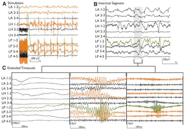
Methods: Intracranial EEG studies were filtered at 500 Hz and sampled at 2,000 Hz. HFOs were visually identified. Twenty patients underwent ES, with gradually increasing currents. Results were interpreted as agreeing or disagreeing with the intracranial study (clinical-EEG seizure onset defined the SOZ). Current thresholds provoking an AD or seizure were correlated with the rate of HFOs of each channel.
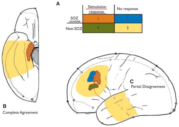
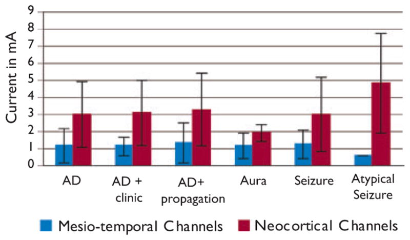
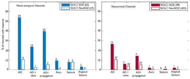
Results: ES provoked a seizure in 12 and ADs in 19 patients. Sixteen patients showed an ES response inside the SOZ, and 10 had additional areas with ADs. The response was more specific for mesiotemporal than for neocortical channels. HFO rates were negatively correlated with thresholds for ES responses; especially in neocortical regions; areas with low threshold and high HFO rate were colocalized even outside the SOZ.
Discussion: Areas showing epileptic HFOs colocalize with those reacting to ES. HFOs may represent a pathologic correlate of regions showing an ES response; both phenomena suggest a more widespread epileptogenicity.
Conflict of interest statement
Disclosure: None of the authors has any conflict of interest to disclose.
Figures
References
-
- Bernier GP, Saint-Hilaire JM, Giard N, Bouvier G, Mercier M. Commentary: intracranial electrical stimulation. In: Engel J Jr, editor. Surgical treatment of epilepsy. Raven press; New York: 1987. pp. 232–334.
-
- Bernier GP, Richer F, Giard N, Bouvier G, Mercier M, Turmel A, Saint-Hilaire JM. Electrical stimulation of the human brain in epilepsy. Epilepsia. 1990;31:513–520. - PubMed
-
- Blume WT, Jones DC, Pathak P. Properties of after-discharges from cortical electrical stimulation in focal epilepsies. Clin Neurophysiol. 2004;115:982–989. - PubMed
-
- Bragin A, Engel J, Jr, Wilson CL, Fried I, Mathern GW. Hippocampal and entorhinal cortex high-frequency oscillations (100–500 Hz) in human epileptic brain and in kainic acid-treated rats with chronic seizures. Epilepsia. 1999;40:127–137. - PubMed
-
- Bragin A, Wilson CL, Staba RJ, Reddick M, Fried I, Engel J., Jr Interictal high-frequency oscillations (80–500 Hz) in the human epileptic brain: entorhinal cortex. Ann Neurol. 2002;52:407–415. - PubMed
Publication types
MeSH terms
Grants and funding
LinkOut - more resources
Full Text Sources
Other Literature Sources
