. 2009 Oct 22:7:62.
doi: 10.1186/1741-7015-7-62.

Genomic and epigenetic evidence for oxytocin receptor deficiency in autism

Affiliations

Genomic and epigenetic evidence for oxytocin receptor deficiency in autism

Simon G Gregory et al. BMC Med. .

Abstract

Background: Autism comprises a spectrum of behavioral and cognitive disturbances of childhood development and is known to be highly heritable. Although numerous approaches have been used to identify genes implicated in the development of autism, less than 10% of autism cases have been attributed to single gene disorders.

Methods: We describe the use of high-resolution genome-wide tilepath microarrays and comparative genomic hybridization to identify copy number variants within 119 probands from multiplex autism families. We next carried out DNA methylation analysis by bisulfite sequencing in a proband and his family, expanding this analysis to methylation analysis of peripheral blood and temporal cortex DNA of autism cases and matched controls from independent datasets. We also assessed oxytocin receptor (OXTR) gene expression within the temporal cortex tissue by quantitative real-time polymerase chain reaction (PCR).

Results: Our analysis revealed a genomic deletion containing the oxytocin receptor gene, OXTR (MIM accession no.: 167055), previously implicated in autism, was present in an autism proband and his mother who exhibits symptoms of obsessive-compulsive disorder. The proband's affected sibling did not harbor this deletion but instead may exhibit epigenetic misregulation of this gene through aberrant gene silencing by DNA methylation. Further DNA methylation analysis of the CpG island known to regulate OXTR expression identified several CpG dinucleotides that show independent statistically significant increases in the DNA methylation status in the peripheral blood cells and temporal cortex in independent datasets of individuals with autism as compared to control samples. Associated with the increase in methylation of these CpG dinucleotides is our finding that OXTR mRNA showed decreased expression in the temporal cortex tissue of autism cases matched for age and sex compared to controls.

Conclusion: Together, these data provide further evidence for the role of OXTR and the oxytocin signaling pathway in the etiology of autism and, for the first time, implicate the epigenetic regulation of OXTR in the development of the disorder.See the related commentary by Gurrieri and Neri: http://www.biomedcentral.com/1741-7015/7/63.


Figures

Figure 1
Figure 1
Graphical representation of the 0.7 Mb heterozygous deletion in 3p25.3 (8,231,927-8,985,513 base pairs) within an individual with autism identified by array comparative genomic hybridization (CGH). The horizontal ideogram represents the chromosome 3 region of interest with the log2 plot below it of array CGH results from the tilepath clones (individual blue circles) on the genomic array. The dotted line at '0' represents copy neutral log2 plot between the cohybridized samples, while the green line represents single copy gain and the red line single copy loss. OXTR and four adjacent genes are contained within a deletion called in Nexus by circular binary segmentation (CBS) [40] (bold black line). Known copy number variants from the Database of Genomic Variants are denoted by horizontal pink bars.
Figure 2
Figure 2
Microsatellite and quantitative real-time polymerase chain reaction (PCR) validation of OXTR deletion. (a) Microsatellite markers within and adjacent to the copy variable region in the OXTR deletion family were assayed to confirm homozygosity and parental transfer of alleles. (b) Normalized real-time PCR results confirm the loss of a single copy of OXTR in both the mother and the proband (marked by a black triangle). Copy number was normalized to RNAseP and controls were used to verify deletion. The 1 × control is a tumor sample with a known chromosome 3 deletion. The 2 × control is an unaffected sample from the copy number variant (CNV) study in this paper that is not copy variable at OXTR.
Figure 3
Figure 3
CpG dinucleotides analyzed within a predicted CpG island in the promoter region of OXTR. Untranslated regions are represented as gray boxes and protein coding exons are white boxes, the site of translation is denoted by a black arrow. Predicted CpG islands are horizontal black bars beneath the gene. The region of interest within the 5' CpG island, MT2 as reported by Kusui et al. [56], is denoted by a dashed line (NCBI36 coordinates). Groups of CpGs sequenced per clone are indicated with brackets. Clear circles with diagonal lines denote untested CpG dinucleotides, filled circles represent region of interest. CpGs of interest are numbered according to translation initiation site of +1. Clear boxes denote CpG dinucleotides that showed no methylation in bisulfite sequencing of subclones; black within boxes denotes methylation of the CpG dinucleotide; F = Father, M = Mother, P = Proband, S = Sibling.
Figure 4
Figure 4
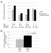
Expression of OXTR in the cortex of male autistic brains is decreased compared to controls. OXTR expression in the cortex was quantitated using quantitative real-time polymerase chain reaction (qRT-PCR) and normalized to PPIA in four cases (autism) and controls matched for age and sex. (a) The expression level of OXTR is decreased in male cases compared to controls and the methylation of site -934 (Me-CpG) correlates with expression in two of the male individuals. (b) As a group, the expression of OXTR in the cortex of the male autistic brain is significantly lower than in controls matched for age and sex. †Paired t test.

References

    1. Yeargin-Allsopp M, Rice C, Karapurkar T, Doernberg N, Boyle C, Murphy C. Prevalence of autism in a US metropolitan area. JAMA. 2003;289:49–55. doi: 10.1001/jama.289.1.49. - DOI - PubMed
    1. Volkmar FR, Szatmari P, Sparrow SS. Sex differences in pervasive developmental disorders. J Autism Dev Disord. 1993;23:579–591. doi: 10.1007/BF01046103. - DOI - PubMed
    1. McLennan JD, Lord C, Schopler E. Sex differences in higher functioning people with autism. J Autism Dev Disord. 1993;23:217–227. doi: 10.1007/BF01046216. - DOI - PubMed
    1. Muhle R, Trentacoste SV, Rapin I. The genetics of autism. Pediatrics. 2004;113:e472–e486. doi: 10.1542/peds.113.5.e472. - DOI - PubMed
    1. Gutierrez GC, Smalley SL, Tanguay PE. Autism in tuberous sclerosis complex. J Autism Dev Disord. 1998;28:97–103. doi: 10.1023/A:1026032413811. - DOI - PubMed

Publication types