. 2009 Dec 20;395(2):255-67.
doi: 10.1016/j.virol.2009.09.026. Epub 2009 Oct 28.

A human coronavirus OC43 variant harboring persistence-associated mutations in the S glycoprotein differentially induces the unfolded protein response in human neurons as compared to wild-type virus

Affiliations

A human coronavirus OC43 variant harboring persistence-associated mutations in the S glycoprotein differentially induces the unfolded protein response in human neurons as compared to wild-type virus

Dominique J Favreau et al. Virology. .

Abstract

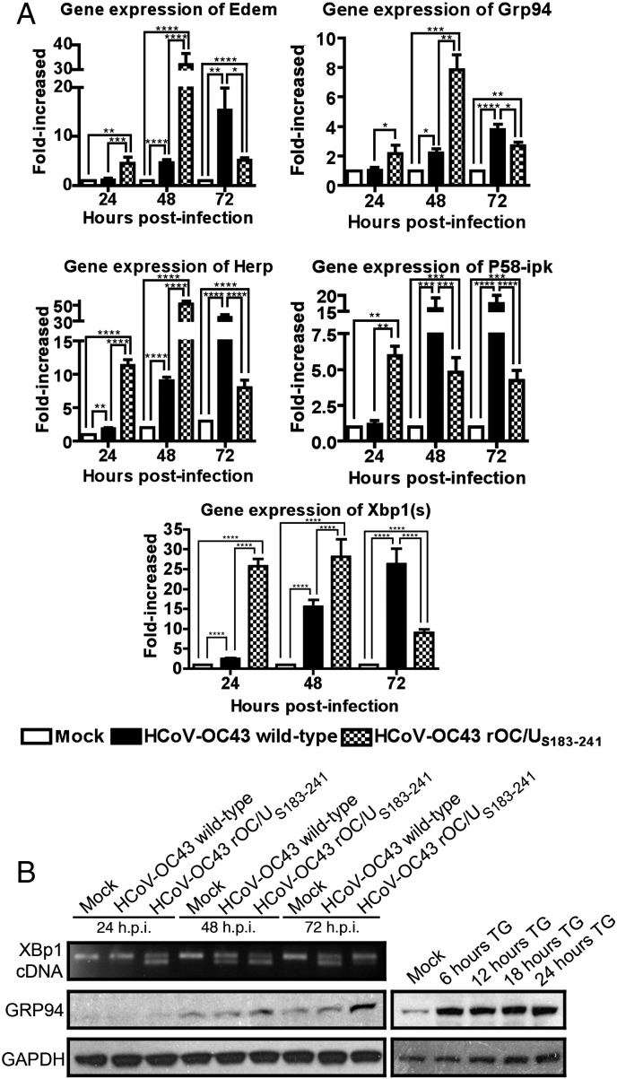
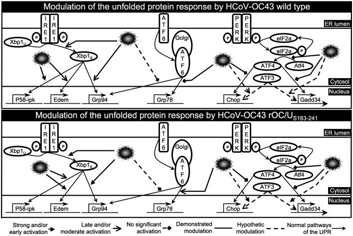
We have reported that human respiratory coronavirus OC43 (HCoV-OC43) is neurotropic and neuroinvasive in humans and mice, and that neurons are the primary target of infection in mice, leading to neurodegenerative disabilities. We now report that an HCoV-OC43 mutant harboring two persistence-associated S glycoprotein point mutations (H183R and Y241H), induced a stronger unfolded protein response (UPR) and translation attenuation in infected human neurons. There was a major contribution of the IRE1/XBP1 pathway, followed by caspase-3 activation and nuclear fragmentation, with no significant role of the ATF6 and eIF2-alpha/ATF4 pathways. Our results show the importance of discrete molecular viral S determinants in virus-neuronal cell interactions that lead to increased production of viral proteins and infectious particles, enhanced UPR activation, and increased cytotoxicity and cell death. As this mutant virus is more neurovirulent in mice, our results also suggest that two mutations in the S glycoprotein could eventually modulate viral neuropathogenesis.


Figures

Fig. 1
Fig. 1
Human differentiated neuronal cell lines were susceptible to a productive HCoV-OC43 infection. (A) Immunofluorescence on cell lines differentiated into human neurons, LA-N-5 (panels a and c) and NT2-N (panels b and d), using specific neuronal markers NFM (panel a) and β-tub isoform III (panel b), or detection of HCoV-OC43 (panels c and d). (B) Production of infectious HCoV‑OC43 particles in supernatants of infected LA-N-5 and NT2-N cell lines differentiated into human neurons.
Fig. 2
Fig. 2
Distribution of the 275 genes which expression was modulated in the human NT2-N neuronal cell line at 24, 48 or 72 h after infection with HCoV-OC43, grouped into nine major cellular functional groups. Neurogenesis/neurotransmission: 29 genes; mobility/adhesion: 24 genes; biosynthesis/degradation: 36 genes; immunity: 15 genes; transcription/replication: 33 genes; cell signaling: 55 genes; cell metabolism: 34 genes; cytoskeleton: 22 genes; apoptosis/cell survival: 27 genes.
Fig. 3
Fig. 3
A recombinant HCoV-OC43 with two point mutations within the S protein showed increased viral replication within infected neurons but no difference in the kinetics of replication in cell culture compared to HCoV-OC43 wild-type. LA-N-5 cells were infected with HCoV-OC43 wild-type or HCoV-OC43 rOC/US183-241 variant for 4, 8, 12, 16, 20, 24, 48 or 72 h. (A) Production of viral infectious particles. Supernatants and cells were separately harvested and titers of extra-and intra-cellular viral infectious particles were assayed. (B) Detection of viral antigens within neurons infected by HCoV-OC43 wild-type or HCoV-OC43 rOC/US183-241. Merged images of immunofluorescence of LA-N-5 differentiated into human neurons infected with HCoV-OC43 wild-type (left panel) or HCoV-OC43 rOC/US183-241 (right panel) showing HCoV-OC43 antigens (green) and DAPI (blue). (C) Kinetics of infection of neurons. Neurons were infected and, at the indicated times post-infection, fixed, permeabilized, labeled with antibody directed against HCoV-OC43 or HCoV-229E (isotype control) and labeled with secondary antibody AlexaFluor 488 anti-mouse. Grey area represent cells labeled with isotype control antibody and white area represent cells labeled with HCoV-OC43 antibody. (D) Percentage of infected neurons. Percentage of cells labeled with HCoV-OC43 antibody were quantified considering the background established with cells labeled with HCoV-229E isotype control antibody.
Fig. 4
Fig. 4
The ATF6 pathway was not activated following infection of human neurons with HCoV-OC43 wild-type and HCoV-OC43 rOC/US183-241 variant. LA-N-5 cells were infected with HCoV-OC43 wild-type or HCoV-OC43 rOC/US183-241 variant for 24, 48 or 72 h, or incubated in presence of 2 μM of thapsigargin for 6, 12, 18 or 24 h. (A) Grp78 gene expression analysis. Total RNA extracted was reverse-transcribed and mRNA expression was evaluated by quantitative PCR compared to mock-infected cells. Gapdh expression was used for normalization. Statistical significance: ⁎p <  0.05; ⁎⁎p <  0.01; ⁎⁎⁎p <  0.0001. (B) GRP78 protein expression analysis. Whole cell lysates were subjected to Western blot analysis using antibody directed against GRP78. GAPDH served as a loading control. Thapsigargin-treated samples served as a positive control.
Fig. 5
Fig. 5
Infection of human neurons with the HCoV-OC43 rOC/US183-241 variant led to a stronger protein translation attenuation, as compared to HCoV-OC43 wild-type. The LA-N-5 cells were infected with HCoV-OC43 wild-type or HCoV-OC43 rOC/US183-241 for 24, 48 or 72 h, or incubated in the presence of 2 μM of thapsigargin for 2 h. (A) eIF2-alpha phosphorylation-state analysis. Whole cell lysates were subjected to Western blot analysis using antibody directed against Ser52-phosphorylated eIF2-alpha. Total eIF2-alpha served as a loading control. Thapsigargin-treated samples served as a positive control. (B and C) Protein synthesis analysis. Cell cultures were starved for cysteine and methionine for 30 min, then incubated with 35S-radiolabeled cysteine/methionine for 15 min and harvested. Whole cell lysates were assayed for cpm (C) counts and resolved on a 4–12% polyacrylamide gel (B). Thapsigargin-treated cells served as a translational shutoff control. (D) Cell viability assay. Cell cultures were incubated with the MTS/PMS solution and absorbance was read at 492 nm. Cell viability is expressed as a relative percentage compared to mock-infected cells. Statistical significance: ⁎p <  0.05; ⁎⁎p <  0.001; ⁎⁎⁎p <  0.0001; ⁎⁎⁎⁎p <  0.000001.
Fig. 6
Fig. 6
Infection of human neurons with the HCoV-OC43 rOC/US183-241 variant strongly induced expression of genes Chop and Gadd34, compared to HCoV-OC43 wild-type. The LA-N-5 cells were infected with HCoV-OC43 wild-type or HCoV-OC43 rOC/US183-241 for 24, 48 or 72 h, or incubated in presence of 2 μM of thapsigargin for 6, 12, 18 or 24 h. (A) Chop and Gadd34 gene expression analysis. Total RNA was extracted and reverse-transcribed and mRNA expression was evaluated by quantitative PCR compared to mock-infected. Gapdh expression was used for normalization. Statistical significance: ⁎p < 0.05; ⁎⁎p < 0.01; ⁎⁎⁎p <  0.001; ⁎⁎⁎⁎p <  0.0001. (B) ATF4, CHOP and GADD34 protein expression analysis. Whole cell lysates were subjected to Western blot analysis using antibody directed against ATF4, CHOP or GADD34. GAPDH served as a loading control. Thapsigargin-treated samples served as a positive control. ⁎Non-specific bands on ATF4 Western blot.
Fig. 7
Fig. 7
The IRE1/XBP1 pathway was rapidly activated following infection of human neurons with the HCoV-OC43 rOC/US183-241 variant, as compared to HCoV-OC43 wild-type. The LA-N-5 cells were infected with HCoV-OC43 wild-type or HCoV-OC43 rOC/US183-241 for 24, 48 or 72 h, or incubated in presence of 2 μM of thapsigargin for 6, 12, 18 or 24 h. (A) Xbp1(s), Xbp1(u)(s), Grp94, Edem, Herp and P58-ipk gene expression analysis. Total RNA was extracted and reverse-transcribed and mRNA expression was evaluated by quantitative PCR compared to mock-infected. Gapdh expression was used for normalization. Statistical significance: ⁎p <  0.05; ⁎⁎p <  0.01; ⁎⁎⁎p <  0.001; ⁎⁎⁎⁎p <  0.0001. (B) GRP94 protein expression analysis. Whole cell lysates were subjected to Western blot analysis using antibody directed against GRP94. GAPDH served as a loading control. Thapsigargin-treated samples served as a positive control.
Fig. 8
Fig. 8
Infection of human neurons by HCoV-OC43 rOC/US183-241 led to a strong activation of caspase-3 and nuclear fragmentation following strong UPR activation. Detection of activation of caspase-3 and nuclear fragmentation within neurons infected by HCoV-OC43 wild-type or HCoV-OC43 rOC/US183-241. (A) Immunofluorescence of LA-N-5 differentiated into human neurons infected with HCoV-OC43 wild-type or HCoV-OC43 rOC/US183-241 showing HCoV-OC43 S protein (red), caspase-3 activation (green), their colocalization in merged image (yellow) and nuclear fragmentation by DAPI (blue). Arrows indicate example of infected cells with activated caspase-3. Arrowheads show example of cells with intense fragmented nucleus. Staurosporine-treated samples served as a positive control. (B) Quantification of cells showing nuclear fragmentation. Data are presented as a percentage of cells with nuclear fragmentation compared to total cells in the field. Each quantification represents the mean of 5 fields containing at least 100 cells. Statistical significance: ⁎p <  0.001.
Fig. 9
Fig. 9
Model depicting the activation of the normal UPR and possible modulation of the different pathways by the HCoV-OC43 S protein in human neuronal cells. The IRE1/XBP1 pathway is strongly and rapidly activated by the HCoV-OC43 rOC/US183-241 mutant while its activation is less intense and slower by HCoV-OC43 ATCC. Target genes of XBP1, e.g. Grp94, P58-ipk and Edem, are expressed following the appearance of the Xbp1S spliced form. The ATF6 pathway is thought to play a minor role, if any, in the UPR activated by both viruses. Indeed, its well known major target gene, Grp78, is merely expressed and no expression upregulation of the GRP78 protein is observed in both infection. The PERK/eIF2α pathway is only transiently activated by both viruses in the first 24 h post-infection although HCoV-OC43 rOC/US183-241 induced a strong translational protein shutoff starting at 48 h post-infection. However, no ATF4 protein was found during the course of infections by both viruses, even though Chop and Gadd34 gene were expressed. This could be explained by the expression of ATF3, also known to regulate their expression. Acronyms. IRE1: inositol-requiring enzyme 1; PERK: PKR-like ER kinase; ATF6: activating transcription factor 6; Xbp1U: X-box binding protein mRNA unspliced form; Xbp1S: X-box binding protein mRNA spliced form; ATF4: activating transcription factor 4; ATF3: activating transcription factor 3; eIF2α: α subunit of eukaryotic translation initiation factor 2.

Similar articles

Cited by

References

    1. Antony J.M., Ellestad K.K., Hammond R., Imaizumi K., Mallet F., Warren K.G., Power C. The human endogenous retrovirus envelope glycoprotein, syncytin-1, regulates neuroinflammation and its receptor expression in multiple sclerosis: a role for endoplasmic reticulum chaperones in astrocytes. J. Immunol. 2007;179:1210–1224. - PubMed
    1. Arbour N., Côté G., Lachance C., Tardieu M., Cashman N.R., Talbot P.J. Acute and persistent infection of human neural cell lines by human coronavirus OC43. J. Virol. 1999;73:3338–3350. - PMC - PubMed
    1. Arbour N., Day R., Newcombe J., Talbot P.J. Neuroinvasion by human respiratory coronaviruses. J. Virol. 2000;74:8913–8921. - PMC - PubMed
    1. Bechill J., Chen Z., Brewer J.W., Baker S.C. Coronavirus infection modulates the unfolded protein response and mediates sustained translational repression. J. Virol. 2008;82:4492–4501. - PMC - PubMed
    1. Bonavia A., Arbour N., Yong V.W., Talbot P.J. Infection of primary cultures of human neural cells by human coronaviruses 229E and OC43. J. Virol. 1997;71:800–806. - PMC - PubMed

Publication types

MeSH terms