Review
. 2009 Dec;30(12):585-91.
doi: 10.1016/j.it.2009.09.002. Epub 2009 Oct 19.

Neonatal immunity: faulty T-helpers and the shortcomings of dendritic cells

Affiliations
Review

Neonatal immunity: faulty T-helpers and the shortcomings of dendritic cells

Habib Zaghouani et al. Trends Immunol. 2009 Dec.

Abstract

Immunity in the newborn is characterized by minimal T helper (Th)1 function but an excess of Th2 activity. Since Th1 lymphocytes are important to counter microbes and Th2 cells favor allergies, the newborn faces susceptibility to microbial infections and allergic reactions. Delayed maturation of certain dendritic cells leads to limited interleukin (IL)-12 production during the neonatal period. The Th2 cytokine locus of neonatal CD4(+) T cells is poised epigenetically for rapid and robust production of IL-4 and IL-13. Together, these circumstances lead to efficient differentiation of Th2 cells and the expression of an IL-4Ralpha/IL-13Ralpha1 heteroreceptor on Th1 cells. Upon re-challenge, Th2 cells rapidly produce IL-4 which utilizes the heteroreceptor to drive apoptosis of Th1 cells, thus yielding the Th2 bias of neonatal immunity.


Figures

Figure 1
Figure 1. Exposure to antigen at birth leads to Th2 responses that preserve tolerance
Early studies showed that when newborns are given antigen (Ag) on the day of birth and challenged with the same Ag as adults, tolerance develops (left). However, if newborn littermates are not given the Ag at birth but are challenged with the same regimen, Th1 responses develop and provide immunity (right). Subsequent studies however showed that the newborns given Ag on the day of birth and challenged with the same Ag as adults do in fact respond but rather produce Th2 responses (left panel). Since Th2 cells do not manifest inflammatory reactions to Ag like the Th1 cells, this response has been historically viewed as tolerance.
Figure 2
Figure 2. Primary neonatal responses are comprised of both Th1 and Th2 cells
Primary neonatal immune responses can be tracked using a neonate-to-neonate T cell receptor (TCR) transgenic adoptive transfer system. T cell receptor (TCR) transgenic T cells specific for ovalbumin (OVA)323-339 peptide are harvested from the spleens of DO11.10 neonatal mice, pooled, and transferred to Balb/c newborns to increase the frequency of responding neonatal cells. The hosts were exposed to Ag shortly thereafter and a clonotype-specific anti-TCR antibody was used to trace the responding cells for analysis of their cytokine responses to determine whether they are Th1- or Th2-type cells. This model has revealed that a balanced Th1:Th2 primary response develops in the neonates but the Th1 cells express the IL-13Rα1 chain.
Figure 3
Figure 3. IL-4 utilizes the IL-4α/IL-13Rα1 heteroreceptor to drive apoptosis of Th1 cells during re-challenge with Ag and biases adult secondary immunity towards Th2 cells
IL-13Rα1 expressed on primary Th1 cells heterodimerizes with IL-4Rα and forms an IL-4α/IL-13Rα1 heteroreceptor. When neonatally primed mice are rechallenged as adults with specific antigen (Ag), IL-4 from Th2 cells binds to the heteroreceptor on Th1 cells and drives their apoptosis resulting in a bias of secondary responses towards Th2 cells.
Figure 4
Figure 4. Neonatal epigenetic readiness sustains rapid production of IL-4
The neonatal Th2 cytokine locus is epigenetically poised for rapid Th2 cytokine production because CNS-1, a key Th2 regulatory region, pre-exists in a relatively hypomethylated (white circles) state in naïve neonatal CD4+ cells. This leads to a default state in which there is rapid production of Th2 cytokines under neutral conditions (i.e. not biased with cytokines and blocking antibodies to produce Th2 cells). In contrast, CNS-1 is hypermethylated (black circles) in naïve adult CD4+ cells and the production of high levels of Th2 cytokines requires longer stimulation times under Th2-promoting conditions. Thus, the pre-existing epigenetic modifications in neonates provide them with the competency to produce high levels of Th2 cytokines under neutral conditions and more rapidly than adult cells, even when the latter are activated under Th2-promoting conditions.
Figure 5
Figure 5. CD8α+CD4 dendritic cell subset reaches optimal frequency on day 6 after birth and produces sufficient IL-12 to suppress IL-13Rα1 up-regulation on neonatal Th1 cells
Most dendritic cells (DCs) in newborn mice spleens are of the CD8αCD4 phenotype. Maturation begins soon after birth and by day 6 both CD8α+CD4 and CD8αCD4+ reach optimal frequency. If antigen (Ag) is given on day 1, the low numbers of Ag presenting CD8α+CD4 DCs, results in very little IL-12 being produced. While this level of IL-12 is sufficient for Th1 differentiation, it is not enough to suppress IL-13Rα1 up-regulation on Th1 cells. By day 6 due to maturation and increase in the CD8α+CD4 DC subset, IL-12 production is augmented and sufficient to suppress IL-13Rα1 up-regulation on Th1 cells.
Figure 6
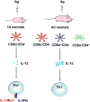
Figure 6. An updated model of neonatal immunity
The frequency of the CD8α+CD4 dendritic cell (DC) subset in the neonatal environment is minimal on the day of birth. Exposure to antigen (Ag) during the neonatal period leads to reduced production of IL-12 by the Ag-presenting CD8α+CD4 DCs which is not sufficient to downregulate IL-13Rα1 expression on the developing primary Th1 cells. As a result, IL-13Rα1 associates with IL-4Rα to form a type II IL-4Rα/IL-13Rα1 heteroreceptor. Upon rechallenge of the adult mice with the same antigen, IL-4 produced by Th2 cells signals through the heteroreceptor and causes apoptosis of Th1 cells. However, by day 6 after birth, DC maturation yields a significant number of CD8a+CD4 DCs which produce sufficient amounts of IL-12 to downregulate IL-13Rα1 expression on Th1 cells. Consequently, IL-4Rα/IL-13Rα1 heteroreceptor formation becomes minimal and IL-4 produced by Th2 cells during re-challenge with antigen can no longer drive apoptosis of Th1 cells and secondary Th1 responses develop.

References

    1. Billingham RE, et al. Actively acquired tolerance of foreign cells. Nature. 1953;172:603–606. - PubMed
    1. Gammon G, et al. Neonatal T cell tolerance to minimal immunogenic peptides is caused by clonal inactivation. Nature. 1986;319:413–415. - PubMed
    1. Clayton JP, et al. Peptide-specific prevention of experimental allergic encephalomyelitis: Neonatal tolerance induced to the dominant T cell determinant of myelin basic protein. J Exp Med. 1989;169:1681–1691. - PMC - PubMed
    1. Lawn J, et al. 4 million neonatal deaths: When? Where? Why? Lancet. 2005;365:9–18. - PubMed
    1. Prescott S, et al. Development of allergen-specific T-cell memory in atopic and normal children. Lancet. 1999;353:196–200. - PubMed

Publication types