. 2009 Dec 18;284(51):35534-42.
doi: 10.1074/jbc.M109.072132.

Mycobacterial cytochrome p450 125 (cyp125) catalyzes the terminal hydroxylation of c27 steroids

Affiliations

Mycobacterial cytochrome p450 125 (cyp125) catalyzes the terminal hydroxylation of c27 steroids

Jenna K Capyk et al. J Biol Chem. .

Abstract

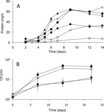
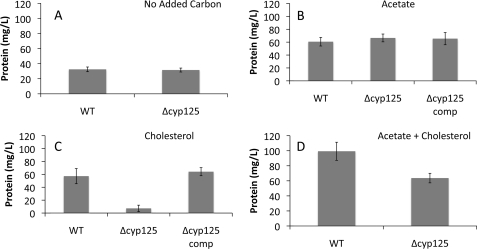
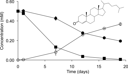
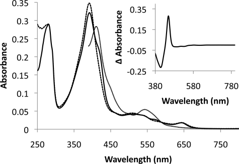
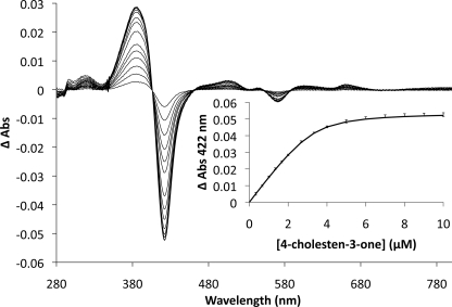
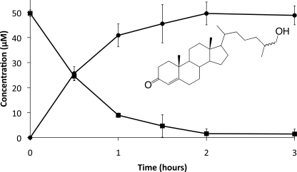
Cyp125 (Rv3545c), a cytochrome P450, is encoded as part of the cholesterol degradation gene cluster conserved among members of the Mycobacterium tuberculosis complex. This enzyme has been implicated in mycobacterial pathogenesis, and a homologue initiates cholesterol catabolism in the soil actinomycete Rhodococcus jostii RHA1. In Mycobacterium bovis BCG, cyp125 was up-regulated 7.1-fold with growth on cholesterol. A cyp125 deletion mutant of BCG did not grow on cholesterol and accumulated 4-cholesten-3-one when incubated in the presence of cholesterol. Wild-type BCG grew on this metabolite. By contrast, a parallel cyp125 deletion mutation of M. tuberculosis H37Rv did not affect growth on cholesterol. Purified Cyp125 from M. tuberculosis, heterologously produced in R. jostii RHA1, bound cholesterol and 4-cholesten-3-one with apparent dissociation constants of 0.20 +/- 0.02 microM and 0.27 +/- 0.05 microm, respectively. When reconstituted with KshB, the cognate reductase of the ketosteroid 9alpha-hydroxylase, Cyp125 catalyzed the hydroxylation of these steroids. MS and NMR analyses revealed that hydroxylation occurred at carbon 26 of the steroid side chain, allowing unambiguous classification of Cyp125 as a steroid C26-hydroxylase. This study establishes the catalytic function of Cyp125 and, in identifying an important difference in the catabolic potential of M. bovis and M. tuberculosis, suggests that Cyp125 may have an additional function in pathogenesis.


Figures

FIGURE 1.
FIGURE 1.
Construction of mycobacterial Δcyp125 mutants. A, genetic organization of the cyp125 locus in WT strains H37Rv and BCG-Pasteur together with the corresponding Δcyp125 mutants. The size of the BamHI fragments as well as the location of the probe used for Southern analysis are indicated. γδres indicates the res-sites of the γδ-resolvase; hyg indicates the hygromycin resistance gene. B, Southern analysis of BamHI-digested genomic DNA from BCG, its Δcyp125 mutant, H37Rv, and its Δcyp125 mutant. Gene deletion was confirmed employing a [α-32P]dCTP-labeled probe hybridizing to the position indicated in A.
FIGURE 2.
FIGURE 2.
Growth of mycobacterial Δcyp125 mutants. A, representative growth curves of WT BCG (solid symbols) and Δcyp125 mutant (open symbols) on basal medium containing no added carbon source (circles), 10 mm acetate (triangles), or 0.5 mm cholesterol (squares). B, growth of WT Mtb H37Rv (solid symbols) and Δcyp125 mutant (open symbols) on basal medium containing no added carbon source (circles) or 0.5 mm cholesterol (squares). Values represent mean of triplicates ± S.D.
FIGURE 3.
FIGURE 3.
Final protein yields of wild-type (BCG), mutant (Δcyp125), and complementation (Δcyp125-C) strains. Cultures were incubated for 14 days in basal medium supplemented with no added substrate (A), 10 mm acetate (B), 0.5 mm cholesterol (C), or 10 mm acetate plus 0.5 mm cholesterol (D). Plotted values represent mean values of 3–6 replicates, and error bars indicate S.E.
FIGURE 4.
FIGURE 4.
Cholesterol transformation by BCG and Δcyp125 cultures. Cholesterol removal (solid symbols) and 4-cholesten-3-one (inset) accumulation (open symbols) is shown for BCG (squares) and Δcyp125 (circles) growing in medium with 10 mm acetate plus 0.5 mm cholesterol. Plotted values are the average of duplicate experiments, and error bars indicate S.E.
FIGURE 5.
FIGURE 5.
Spectra of Cyp125. The spectra of 3.4 μm Cyp125 as isolated with oxidized heme iron (solid line), upon addition of 10 μm 4-cholesten-3-one (dashed line) and subsequent reduction (dotted line) are shown. The reduced CO-difference spectrum is shown in the inset. Spectra were collected at 25 °C in 0.1 mm potassium phosphate buffer, pH 7.0.
FIGURE 6.
FIGURE 6.
Binding of 4-cholesten-3-one to Cyp125. Difference spectra of 3.3 μm Cyp125 upon the addition of 0.33 to 10 μm of 4-cholesten-3-one are overlaid. The resultant binding curve is inset with error bars representing the S.E. calculated from triplicate data; the solid line represents the best fit of a quadratic binding equation to the data as determined using the program R, with fitted parameters: KD = 0.27 ± 0.05 μm, [Cyp125] = 3.3 ± 0.2 μm, and ΔA422Max = 0.055 ± 0.001. 4-Cholesten-3-one was prepared as an aqueous solution in 10% 2-hydroxypropyl-β-cyclodextrin.
FIGURE 7.
FIGURE 7.
Turnover of 4-cholesten-3-one by Cyp125. Depletion of 4-cholesten-3-one and accumulation of 26-hydroxy-4-cholesten-3-one (inset) are represented by squares and circles, respectively. Reactions were carried out in air-saturated 0.1 mm potassium phosphate, pH 7.0, containing 900 μm NADH, 50 μm 4-cholesten-3-one, 1.5 μm KshB, and 1.6 μm Cyp125.

References

    1. World Health Organization (2008) Anti-tuberculosis Drug Resistance in the World, Report 4, pp. 14–21, World Health Organization, Geneva
    1. Munro A. W., McLean K. J., Marshall K. R., Warman A. J., Lewis G., Roitel O., Sutcliffe M. J., Kemp C. A., Modi S., Scrutton N. S., Leys D. (2003) Biochem. Soc. Trans. 31, 625–630 - PubMed
    1. van der Geize R., Yam K., Heuser T., Wilbrink M. H., Hara H., Anderton M. C., Sim E., Dijkhuizen L., Davies J. E., Mohn W. W., Eltis L. D. (2007) Proc. Natl. Acad. Sci. U.S.A. 104, 1947–1952 - PMC - PubMed
    1. Mohn W. W., van der Geize R., Stewart G. R., Okamoto S., Liu J., Dijkhuizen L., Eltis L. D. (2008) J. Biol. Chem. 283, 35368–35374 - PMC - PubMed
    1. Lack N. A., Yam K., Lowe E. D., Horsman G. P., Owen R. L., Sim E., Eltis L. (2009) J. Biol. Chem., in press - PMC - PubMed

Publication types

MeSH terms

Grants and funding