. 2010 Feb;82(2):363-72.
doi: 10.1095/biolreprod.109.078550. Epub 2009 Oct 21.

Isolation, characterization, and culture of human spermatogonia

Affiliations

Isolation, characterization, and culture of human spermatogonia

Zuping He et al. Biol Reprod. 2010 Feb.

Abstract

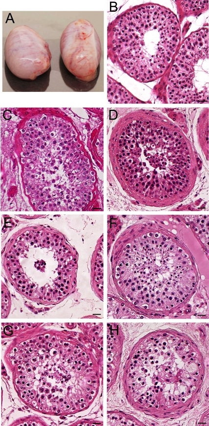
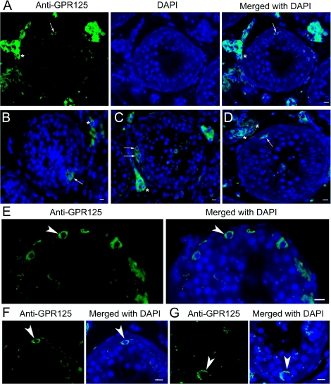
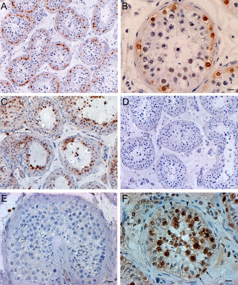
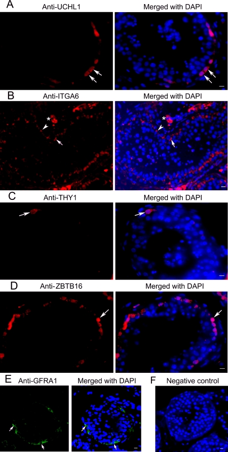
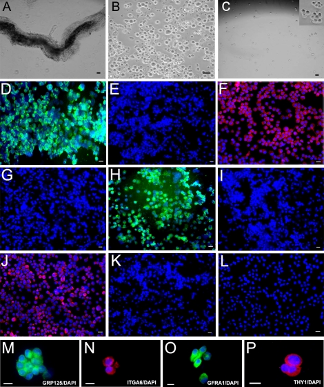
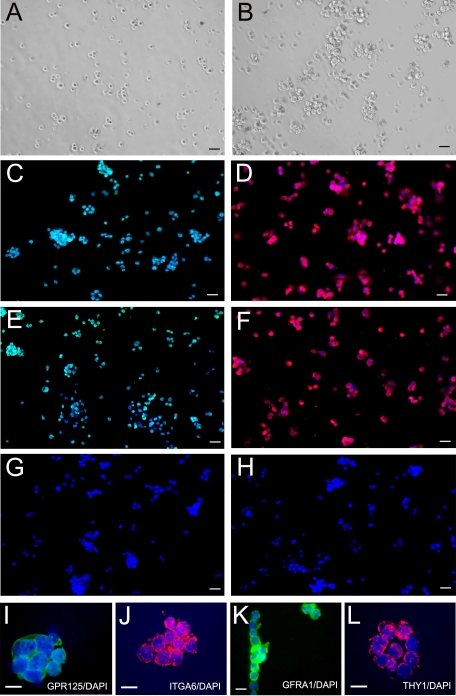
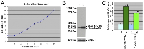
This study was designed to isolate, characterize, and culture human spermatogonia. Using immunohistochemistry on tubule sections, we localized GPR125 to the plasma membrane of a subset of the spermatogonia. Immunohistochemistry also showed that MAGEA4 was expressed in all spermatogonia (A(dark), A(pale), and type B) and possibly preleptotene spermatocytes. Notably, KIT was expressed in late spermatocytes and round spermatids, but apparently not in human spermatogonia. UCHL1 was found in the cytoplasm of spermatogonia, whereas POU5F1 was not detected in any of the human germ cells. GFRA1 and ITGA6 were localized to the plasma membrane of the spermatogonia. Next, we isolated GPR125-positive spermatogonia from adult human testes using a two-step enzymatic digestion followed by magnetic-activated cell sorting. The isolated GPR125-positive cells coexpressed GPR125, ITGA6, THY1, and GFRA1, and they could be cultured for short periods of time and exhibited a marked increase in cell numbers as shown by a proliferation assay. Immunocytochemistry of putative stem cell genes after 2 wk in culture revealed that the cells were maintained in an undifferentiated state. MAPK1/3 phosphorylation was increased after 2 wk of culture of the GPR125-positive spermatogonia compared to the freshly isolated cells. Taken together, these results indicate that human spermatogonia share some but not all phenotypes with spermatogonial stem cells (SSCs) and progenitors from other species. GPR125-positive spermatogonia are phenotypically putative human SSCs and retain an undifferentiated status in vitro. This study provides novel insights into the molecular characteristics, isolation, and culture of human SSCs and/or progenitors and suggests that the MAPK1/3 pathway is involved in their proliferation.


Figures

FIG. 1.
FIG. 1.
Morphology of the donor testes. A) The human testes from the 16-yr-old donor. Each testis weighed ∼14 g and was about 4.1 × 2 × 2.5 cm. BH) H&E staining shows the morphology of the human testes from the five donors. Spermatogenesis appeared normal in all donors, although at times the fixation was not ideal in all regions of the testes. Bars = 20 μm (BH).
FIG. 2.
FIG. 2.
Immunohistochemistry shows GPR125 expression in human testes. AG) GPR125 was localized at plasma membrane of a subset of human spermatogonia (arrows in AD; arrowheads in EG) adjacent to the basement membrane of the seminiferous epithelium. Representative pictures show that very few spermatogonia (arrows and arrowheads) were positive for GPR125 in each seminiferous tubule cross section. GPR125 staining was also observed in Leydig cells (asterisks). Bars = 10 μm.
FIG. 3.
FIG. 3.
Immunohistochemistry reveals the expression of MAGEA4, KIT, POU5F1, and PCNA in human testes. A, B) MAGEA4 was expressed in all spermatogonia (Adark, Apale, and type B spermatogonia) and possibly in preleptotene spermatocytes. C) KIT was expressed in late-stage spermatocytes and round spermatids but not in other germ cells or Sertoli cells. D) Replacement of primary antibody with PBS served as negative control and no staining was observed. E) POU5F1 was undetected in human germ cells but was found occasionally in the nucleus of interstitial cells (asterisk). F) PCNA was expressed in some proliferating spermatogonia (arrows), possibly the renewing Apale, along the basement membrane of the seminiferous tubules and proliferating spermatocytes including pachytene spermatocytes, but not in nonproliferating spermatogonia (possibly the reserve Adark) and preleptotene spermatocytes (arrowheads). Some proliferating interstitial cells (asterisks) were also positive for PCNA. Bars = 20 μm (A, CF), 10 μm (B).
FIG. 4.
FIG. 4.
Immunohistochemistry shows expression of UCHL1, ITGA6, THY1, ZBTB16, and GFRA1 in human testes. A) Expression of UCHL1 was observed in the cytoplasm of the spermatogonia (arrows). B) Expression of ITGA6 was seen in the plasma membrane of spermatogonia (arrows). Often rows of spermatogonia along the basement membrane were stained (arrows). ITGA6 staining was also observed in Sertoli cells (arrowheads) and Leydig cells (asterisks). C) THY1 was expressed in a subpopulation of spermatogonia (arrows). D) ZBTB16 was localized to human spermatogonia (arrows) lying at the basement membrane. E) Expression of GFRA1 was found in the plasma membrane of the spermatogonia (arrows). F) Replacement of primary antibody with PBS served as negative control and no staining was observed. Bars = 10 μm.
FIG. 5.
FIG. 5.
Isolation and characterization of the GPR125-positive spermatogonia from human testis (freshly isolated). A) Seminiferous tubules were isolated from human testes using mechanical dissociation and a one-step enzymatic digestion. B) Human germ cells were obtained using a second enzymatic digestion and different plating. C) GPR125-positive spermatogonia were obtained using MACS with an antibody to GPR125. The inset in C is a high-magnification view showing the GPR125-positive spermatogonia. Images in A, B, and C are phase contrast. After MACS, immunocytochemical analysis showed the expression of GPR125 (D), ITGA6 (F), GFRA1 (H), and THY1 (J), in the isolated GPR125-positive spermatogonia. MP) High magnification showed that freshly isolated GPR125- positive spermatogonia expressed GPR125 (M), ITGA6 (N), GFRA1 (O), and THY1 (P). No staining was observed in GPR125-negative male germ cells using antibody to GPR125 (E), ITGA6 (G), GFRA1 (I), or THY1 (K). L) Replacement of primary antibody with PBS in isolated GPR125-positive spermatogonia served as a negative control. Bars = 10 μm (A, C, D, F, H, J, LP), 20 μm (B, E, G, I, and K).
FIG. 6.
FIG. 6.
Culture for 14 days and characterization of human GPR125-positive spermatogonia. A) GPR125-positive spermatogonia were cultured in StemPro medium supplemented with GDNF, soluble GFRA1, NUDT6 LIF, EGF, Nodal, and TGFB (cells shown at day 1). B) GPR125-positive spermatogonia were cultured in StemPro medium supplemented with growth factors as mentioned above (cells shown at day 14). Panels A and B are phase contrast images. After culture for 14 days, immunocytochemical analysis showed the expression of GPR125 (C), ITGA6 (D), GFRA1 (E), and THY1 (F) in the cultured GPR125-positive spermatogonia. G) Replacement of primary antibody with PBS served as a negative control. H) Replacement of primary antibody with normal rabbit IgG served as another negative control. IL) High magnification shows that cultured GPR125-positive spermatogonia expressed GPR125 (I), ITGA6 (J), GFRA1 (K), and THY1 (L). Bars = 20 μm (AH), 10 μm (IL).
FIG. 7.
FIG. 7.
Cell number increased and MAPK1/3 phosphorylation was activated in human GPR125-positive spermatogonia after culture for 14 days. A) Proliferation assay showed a cell growth curve of human GPR125-positive spermatogonia. B) Western blots revealed that the phosphorylation of MAPK1/3 (upper panel) was activated in human GPR125-positive spermatogonia after culture for 14 days with a number of growth factors compared to the freshly isolated GPR125-positive spermatogonia. MAPK1 was used as a loading control for total proteins (lower panel). C) Relative levels of MAPK1/3 phosphorylation in the cultured GPR125-positive spermatogonia compared to the freshly isolated GPR125-positive spermatogonia (designated as 1) after normalization to the signal obtained with MAPK1. Statistically significant differences (P < 0.05) between the cultured and fresh GPR125-positive spermatogonia are indicated by asterisks. B and C) 1, freshly isolated GPR125-positive spermatogonia; 2, the cultured GPR125-positive spermatogonia.

References

    1. Naughton CK, Jain S, Strickland AM, Gupta A, Milbrandt J.Glial cell-line derived neurotrophic factor-mediated RET signaling regulates spermatogonial stem cell fate. Biol Reprod 2006; 74: 314–321. - PubMed
    1. Hofmann MC, Braydich-Stolle L, Dym M.Isolation of male germ-line stem cells; influence of GDNF. Dev Biol 2005; 279: 114–124. - PMC - PubMed
    1. Meng X, Lindahl M, Hyvonen ME, Parvinen M, de Rooij DG, Hess MW, Raatikainen-Ahokas A, Sainio K, Rauvala H, Lakso M, Pichel JG, Westphal H, et al. Regulation of cell fate decision of undifferentiated spermatogonia by GDNF. Science 2000; 287: 1489–1493. - PubMed
    1. He Z, Jiang J, Hofmann MC, Dym M.Gfra1 silencing in mouse spermatogonial stem cells results in their differentiation via the inactivation of RET tyrosine kinase. Biol Reprod 2007; 77: 723–733. - PMC - PubMed
    1. Yoshinaga K, Nishikawa S, Ogawa M, Hayashi S, Kunisada T, Fujimoto T, Nishikawa S.Role of c-kit in mouse spermatogenesis: identification of spermatogonia as a specific site of c-kit expression and function. Development 1991; 113: 689–699. - PubMed

Publication types

MeSH terms