. 2009 Dec;102(6):3740-52.
doi: 10.1152/jn.00544.2009. Epub 2009 Oct 21.

The subthalamic nucleus in primary dystonia: single-unit discharge characteristics

Affiliations

The subthalamic nucleus in primary dystonia: single-unit discharge characteristics

Lauren E Schrock et al. J Neurophysiol. 2009 Dec.

Abstract

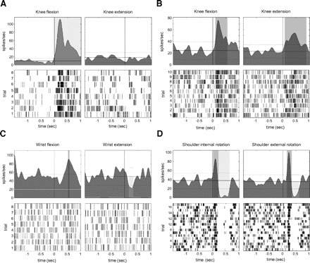
Most models of dystonia pathophysiology predict alterations of activity in the basal ganglia thalamocortical motor circuit. The globus pallidus interna (GPi) shows bursting and oscillatory neuronal discharge in both human dystonia and in animal models, but it is not clear which intrinsic basal ganglia pathways are implicated in this abnormal output. The subthalamic nucleus (STN) receives prominent excitatory input directly from cortical areas implicated in dystonia pathogenesis and inhibitory input from the external globus pallidus. The goal of this study was to elucidate the role of the STN in dystonia by analyzing STN neuronal discharge in patients with idiopathic dystonia. Data were collected in awake patients undergoing microelectrode recording for implantation of STN deep brain stimulation electrodes. We recorded 62 STN neurons in 9 patients with primary dystonia. As a comparison group, we recorded 143 STN neurons in 20 patients with Parkinson's disease (PD). Single-unit activity was discriminated off-line by principal component analysis and evaluated with respect to discharge rate, bursting, and oscillatory activity. The mean STN discharge rate in dystonia patients was 26.3 Hz (SD 13.6), which was lower than that in the PD patients (35.6 Hz, SD 15.2), but higher than published values for subjects without basal ganglia dysfunction. Oscillatory activity was found in both disorders, with a higher proportion of units oscillating in the beta range in PD. Bursting discharge was a prominent feature of both dystonia and PD, whereas sensory receptive fields were expanded in PD compared with dystonia. The STN firing characteristics, in conjunction with those previously published for GPi, suggest that bursting and oscillatory discharge in basal ganglia output may be transmitted via pathways involving the STN and provide a pathophysiologic rationale for STN as a surgical target in dystonia.


Figures

Fig. 1.
Fig. 1.
Examples of single-unit activity from a typical subthalamic nucleus (STN) neuron in a patient with cranial-cervical dystonia (left column) and a patient with the akinetic-rigid subtype of Parkinson's disease (PD, right column). A: neuronal recordings. A 2-s interval is shown. B: interspike interval (ISI) histograms, bin size of 1 ms. Inset: expanded timescale demonstrating the absence of ISIs of <3-ms duration, consistent with the neuronal refractory period. C: raster diagrams showing bursting discharge. Bursts as defined by the Poisson “surprise” method (surprise value >5) are labeled with a black bar above spikes that constitute a burst. Note the higher proportion of bursts per total number of spikes shown in the dystonia neuron (0.40 vs. 0.26 in the PD neuron). Consecutive rows (3 s of data per row) from bottom to top represent continuous 36-s recordings. D: autocorrelograms. The right autocorrelogram shows oscillatory activity of about 11 Hz. The unit on the left was not found to have significant oscillations.
Fig. 2.
Fig. 2.
Histograms of spontaneous STN discharge rates in PD (black bars) and dystonia (gray bars). Bin size is 10 Hz. x-axis labels indicate the bin midpoint (i.e., 5 refers to 0–9.99, 15 refers to 10–19.99, 25 to 20–20.99, etc.). Bars are slightly offset for visual clarity. STN neuronal discharge rate was significantly lower in dystonia patients than that in PD patients (P < 0.001).
Fig. 3.
Fig. 3.
A: distribution of frequencies of significant oscillations in spontaneous STN activity in dystonia (top graph) and PD (bottom graph). Bins are left-inclusive, except for the first bin, which starts at 0.4 Hz since the lower limit of oscillatory activity was 0.49 Hz. (i.e., the bins represent 0.4–2.99, 3–7.99, 8–12.99, 13–29.99, 30–59.99, 60–99.99, and 100–199.99 Hz). Several cells oscillated at more than one frequency (see results). There was a greater incidence of oscillatory activity in low-frequency bands (3–30 Hz) in PD than that in dystonia (P = 0.01). Oscillatory activity in the beta band (13–30 Hz) was found only in PD (P = 0.03). B, top: the power spectra of spike trains generated by the global spike shuffling method between 0 and 30 Hz for a patient with the akinetic-rigid subtype of PD. This is the same PD cell displayed in Fig. 1. There is a single significant peak at 11 Hz. Middle: the power spectra between 0 and 30 Hz for a patient with cranial-cervical dystonia. There is a single significant peak at 3.9 Hz. Bottom: the power spectra between 0 and 115 Hz for a patient with akinetic-rigid PD. There are 2 significant peaks at 3.4 and 90.3 Hz.
Fig. 4.
Fig. 4.
Representative STN neuronal responses to passive limb movement in dystonia and PD. For each example, neuronal responses for reciprocal movements are shown in left and right columns. For each neuron, top panel shows a mean spike density function centered at the initial deflection in limb acceleration (time = 0 s). The dotted line shows the thresholds for significant changes in mean discharge rate. The bottom panel shows a raster display of neuronal responses to each individual trial. A: neuronal response to passive knee movement in a dystonia patient: flexion (left) evoked a brisk increase response, but extension (right) resulted in no appreciable change in firing. B: neuronal response to passive knee movement in a patient with PD: flexion (left) and extension (right) both resulted in firing increases. C: neuronal response to passive wrist movement in a patient with PD: flexion (left) resulted in increased firing and extension (right) evoked a decrease in firing. D: neuronal response to passive shoulder movement in a patient with PD: both external rotation (left) and internal rotation (right) evoked a brisk biphasic response of increased neuronal firing followed by decreased firing.

References

    1. Albin RL, Young AB, Penney JB. The functional anatomy of basal ganglia disorders. Trends Neurosci 12: 366–375, 1989 - PubMed
    1. Asanuma K, Ma Y, Okulski J, Dhawan V, Chaly T, Carbon M, Bressman SB, Eidelberg D. Decreased striatal D2 receptor binding in non-manifesting carriers of the DYT1 dystonia mutation. Neurology 64: 347–349, 2005 - PubMed
    1. Bergman H, Wichmann T, Karmon B, DeLong MR. The primate subthalamic nucleus. II. Neuronal activity in the MPTP model of parkinsonism. J Neurophysiol 72: 507–520, 1994 - PubMed
    1. Berman BD, Starr PA, Marks WJ, Jr, Ostrem JL. Induction of bradykinesia with pallidal deep brain stimulation in patients with cranial-cervical dystonia. Stereotact Funct Neurosurg 87: 37–44, 2009 - PMC - PubMed
    1. Bevan MD, Hallworth NE, Baufreton J. GABAergic control of the subthalamic nucleus. Prog Brain Res 160: 173–188, 2007 - PubMed