. 2009 Dec;20(24):5250-9.
doi: 10.1091/mbc.e09-09-0777.

Small Cajal body-specific RNAs of Drosophila function in the absence of Cajal bodies

Affiliations

Small Cajal body-specific RNAs of Drosophila function in the absence of Cajal bodies

Svetlana Deryusheva et al. Mol Biol Cell. 2009 Dec.

Abstract

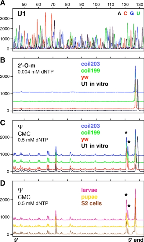
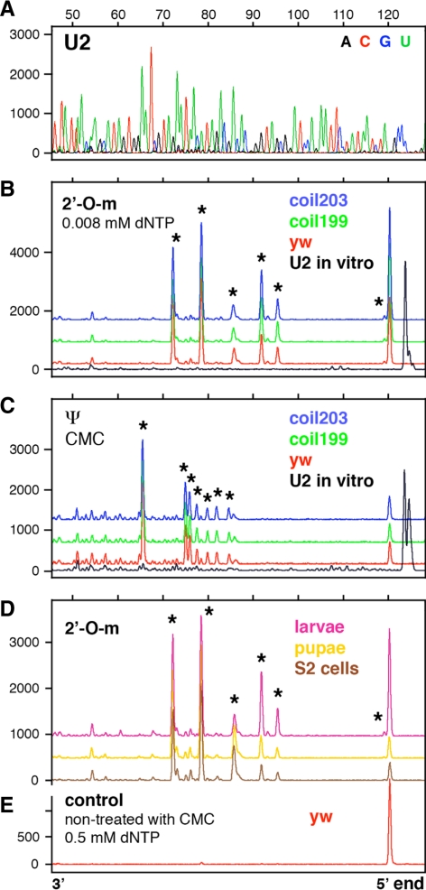
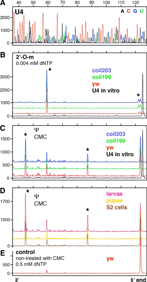
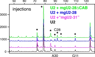
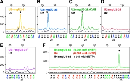
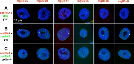
During their biogenesis small nuclear RNAs (snRNAs) undergo multiple covalent modifications that require guide RNAs to direct methylase and pseudouridylase enzymes to the appropriate nucleotides. Because of their localization in the nuclear Cajal body (CB), these guide RNAs are known as small CB-specific RNAs (scaRNAs). Using a fluorescent primer extension technique, we mapped the modified nucleotides in Drosophila U1, U2, U4, and U5 snRNAs. By fluorescent in situ hybridization (FISH) we showed that seven Drosophila scaRNAs are concentrated in easily detectable CBs. We used two assays based on Xenopus oocyte nuclei to demonstrate that three of these Drosophila scaRNAs do, in fact, function as guide RNAs. In flies null for the CB marker protein coilin, CBs are absent and there are no localized FISH signals for the scaRNAs. Nevertheless, biochemical experiments show that scaRNAs are present at normal levels and snRNAs are properly modified. Our experiments demonstrate that several scaRNAs are concentrated as expected in the CBs of wild-type Drosophila, but they function equally well in the nucleoplasm of mutant flies that lack CBs. We propose that the snRNA modification machinery is not limited to CBs, but is dispersed throughout the nucleoplasm of cells in general.


Figures

Figure 1.
Figure 1.
Mapping of 2′-O-methyl groups and pseudouridines in Drosophila U1 snRNA by primer extension. Terminations of the primer extension reaction occur one nucleotide downstream of the actual modification and appear as peaks above the baseline. The strong peak at the 5′ end represents the end of the template molecule. The X-axis shows fragment length in nucleotides determined by the migration of size standards. The Y-axis shows peak intensities in arbitrary units (traces are shifted vertically to avoid overlap). Details are given in Materials and Methods. (A) Sequencing reaction run on in vitro–transcribed U1 snRNA allows the position of terminations to be determined. (B) There are no detectable methylation peaks in U1 snRNA derived from adult control flies (y w), two coilin-null mutants (coil199 and coil203), or in vitro–transcribed U1 snRNA. (C) There are two strong pseudouridine peaks near the 5′ end of U1 snRNA from adult y w and mutant flies (stars at Ψ5 and Ψ6), but not in the control in vitro–transcribed U1 snRNA (black). Multiple small peaks in all the traces represent terminations caused by uridine itself. (D) U1 snRNA derived from S2 cells, larvae, and pupae shows the same two pseudouridine peaks as U1 derived from adult flies (stars at Ψ5 and Ψ6). Note the absence of pseudouridylation at position U22 in all samples.
Figure 2.
Figure 2.
Mapping of 2′-O-methyl groups and pseudouridines in Drosophila U2 snRNA. Conditions as in Figure 1. (A) Sequencing reaction on in vitro–transcribed U2 snRNA. (B) Six 2′-O-methyl groups are detectable in U2 snRNA derived from y w and coilin-null mutant flies (stars at Am1, Gm25, Cm28, Gm34, Cm41, and Um48). (C) Seven pseudouridine modifications are detectable in U2 snRNA derived from y w and coilin-null flies (stars at Ψ35, Ψ38, Ψ40, Ψ42, Ψ44, Ψ45, and Ψ55). (D) U2 snRNA derived from S2 cells, larvae, and pupae shows the same six 2′-O-methyl peaks as U2 derived from adult flies (stars at Am1, Gm25, Cm28, Gm34, Cm41, and Um48). (E) U2 snRNA from y w flies shows no peaks when treated with high concentration of dNTP during the extension reaction. Note the absence of methylation at A31 in all samples.
Figure 3.
Figure 3.
Mapping of 2′-O-methyl groups and pseudouridines in Drosophila U4 snRNA. Conditions as in Figure 1. (A) Sequencing reaction on in vitro–transcribed U4 snRNA. (B) Two 2′-O-methyl groups are detectable in U4 snRNA derived from y w and coilin-null mutant flies (stars at Am1 and Am65). (C) Two pseudouridine modifications are detectable in U4 snRNA derived from y w and coilin-null flies (stars at Ψ36 and Ψ79). The prominent peak at A65 (no star) is due to methylation at this site, which can cause termination of the extension reaction in CMC-treated RNA. (D) U4 snRNA derived from S2 cells, larvae, and pupae shows the same two pseudouridine peaks as U4 derived from adult flies (stars at Ψ36 and Ψ79). (E) U4 snRNA from y w flies shows one tiny peak (Am65) when treated with high concentration of dNTP during the extension reaction. Note the absence of pseudouridine at U59 in all samples.
Figure 4.
Figure 4.
Modification of Drosophila U2 snRNA injected into Xenopus oocytes. When U2 was injected alone, it became methylated in the pattern expected for Xenopus U2 (black trace); that is, at sites common to the two species (stars at Cm25, Gm34, Cm41, and Um48), at Xenopus-specific sites (stars at Gm11 and Am30), but not at a Drosophila-specific site (star at Cm28). When U2 was coinjected with the Drosophila guide for C28 (mgU2-28), an extra peak corresponding to methylation at C28 was observed (blue trace). Methylation at C28 was also observed when the CAB box of its guide RNA was mutated (green trace; mgU2-28ΔCAB). Coinjection of U2 and the putative guide “mgU2-31” did not change the modification pattern (purple trace). If this guide RNA were functional, an additional peak at A31 would be present.
Figure 5.
Figure 5.
Modification of Drosophila U2 snRNA in RNA-depleted Xenopus GV extract. In each case in vitro–transcribed U2 snRNA was added to the extract along with a putative in vitro–transcribed guide RNA. (A) Addition of guide RNA mgU2-41 resulted in a peak of methylation at C41 (yellow trace, star) that was higher than in the control without the guide (black trace). (B) Guide RNA mgU2-28 produced a strong methylation peak at C28 (blue trace, star). (C) Methylation at C28 was also observed when the CAB box of guide RNA mgU2-28 was mutated (mgU2-28ΔCAB, green trace, star). (D) The putative guide RNA mgU2-25 had no effect on methylation (red trace). (E) The putative guide RNA “mgU2-31” had no effect on methylation (purple trace). (F) Modification of Drosophila U4 snRNA in RNA-depleted Xenopus GV extract. Each trace represents a primer extension reaction in which in vitro–transcribed U4 snRNA was added to the extract. Addition of guide RNA mgU4-65 resulted in a peak of methylation at A65 (green trace, star). No peak was seen in the control without the guide (red trace) or in a reaction with the guide but carried out at high concentration of dNTPs (black trace).
Figure 6.
Figure 6.
Northern blots of Drosophila scaRNAs. For each scaRNA there are three lanes that contain total RNA from adult y w, coil199, and coil203 flies, respectively. Both coil199 and coil203 are coilin-null mutants that have no detectable CBs in their cells; y w flies serve as controls. In each case the abundance of the scaRNA is approximately the same for mutant and control flies. Seven Northern blots were run separately with their own size markers. Images of the autoradiographs were grouped into one figure with a common size scale in nucleotides.
Figure 7.
Figure 7.
Confocal images of Malpighian tubule nuclei after two-color FISH for scaRNAs and their cognate snRNAs. The three images in each column show nuclei hybridized with a probe for the scaRNA listed at the top of the column (red probe). The six nuclei in each row were hybridized with U85 scaRNA (A) or the cognate snRNA for the guide RNA (B and C). For instance, the first nucleus in row A was hybridized with probes for mgU2-25 scaRNA (red) and U85 scaRNA (green), whereas the last nucleus in row B was hybridized with probes for mgU5-42 (red) and U5 snRNA (green). Nuclei in rows A and B are from control y w flies and show an easily detectable CB that is labeled with both probes. Nuclei in row C are from coilin-null flies in which a CB is not detectable. All nuclei are stained with the DNA-specific dye DAPI (blue).

References

    1. Andrade L.E.C., Chan E.K.L., Raska I., Peebles C. L., Roos G., Tan E. M. Human autoantibody to a novel protein of the nuclear coiled body: immunological characterization and cDNA cloning of p80-coilin. J. Exp. Med. 1991;173:1407–1419. - PMC - PubMed
    1. Bellini M., Gall J. G. Coilin can form a complex with the U7 small nuclear ribonucleoprotein. Mol. Biol. Cell. 1998;9:2987–3001. - PMC - PubMed
    1. Beumer K. J., Trautman J. K., Bozas A., Liu J.-L., Rutter J., Gall J. G., Carroll D. Efficient gene targeting in Drosophila by direct embryo injection with zinc-finger nucleases. Proc. Natl. Acad. Sci. USA. 2008;105:19821–19826. - PMC - PubMed
    1. Carmo-Fonseca M., Tollervey D., Barabino S.M.L., Merdes A., Brunner C., Zamore P. D., Green M. R., Hurt E., Lamond A. I. Mammalian nuclei contain foci which are highly enriched in components of the pre-mRNA splicing machinery. EMBO J. 1991;10:195–206. - PMC - PubMed
    1. Collier S., Pendle A., Boudonck K., van Rij T., Dolan L., Shaw P. A distant coilin homologue is required for the formation of Cajal bodies in Arabidopsis. Mol. Biol. Cell. 2006;17:2942–2951. - PMC - PubMed

Publication types