. 2009 Oct 21;29(42):13182-9.
doi: 10.1523/JNEUROSCI.2624-09.2009.

Exaggerated response of a vasopressin-enhanced green fluorescent protein transgene to nociceptive stimulation in the rat

Affiliations

Exaggerated response of a vasopressin-enhanced green fluorescent protein transgene to nociceptive stimulation in the rat

Hitoshi Suzuki et al. J Neurosci. .

Abstract

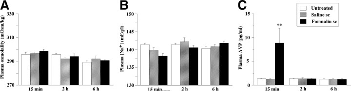
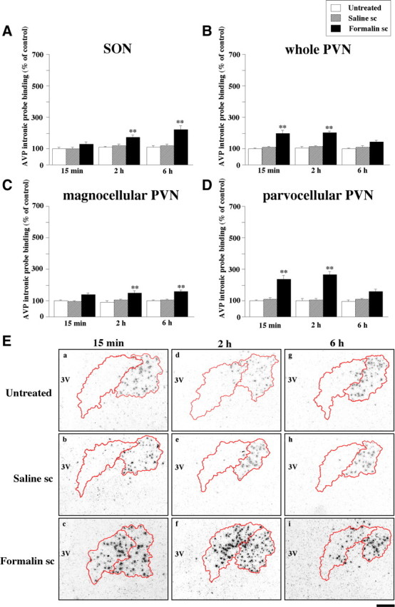
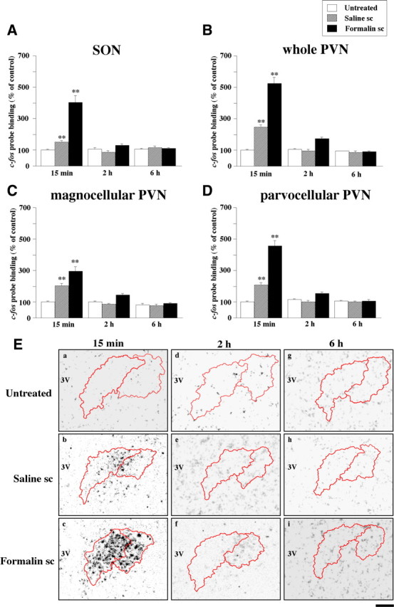
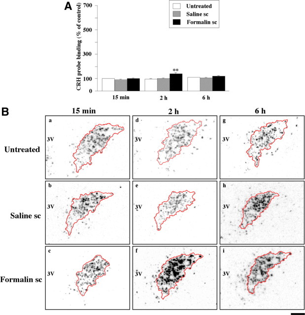
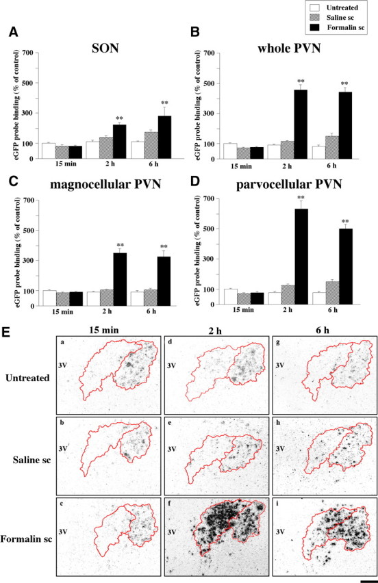
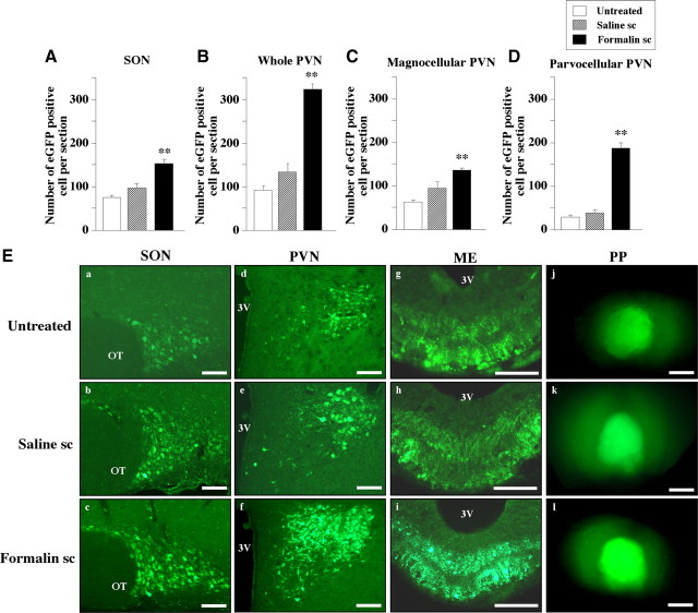
Nociceptive stimulation elicits neuroendocrine responses such as arginine vasopressin (AVP) release as well as activation of the hypothalamo-pituitary-adrenal axis. We have generated novel transgenic rats expressing an AVP-enhanced green fluorescent protein (eGFP) fusion gene, and we examined the effects of nociceptive stimulation on transgene expression in the hypothalamus after subcutaneous injection of saline or formalin into the bilateral hindpaws in these rats. We have assessed (1) AVP levels in plasma and the changes of eGFP mRNA and AVP heteronuclear RNA (hnRNA) in the supraoptic nucleus (SON) and the paraventricular nucleus (PVN) using in situ hybridization histochemistry, (2) gene expression changes in distinct magnocellular and parvocellular divisions of the PVN, (3) eGFP fluorescence in the SON, the PVN, the median eminence (ME), and the posterior pituitary gland (PP). Plasma AVP levels were significantly increased 15 min after formalin injection. In the same time period, the AVP hnRNA levels in the PVN were increased, especially in the parvocellular division of the PVN in formalin-injected rats. In the same region, eGFP mRNA levels after formalin injection were also significantly increased to a much greater extent than those of AVP hnRNA. The eGFP fluorescence in the SON, the PVN, the ME, and the PP was markedly increased in formalin-injected rats and especially increased in the parvocellular divisions of the PVN. Together, our results demonstrate robust and rapid changes in the expression of the AVP-eGFP transgene in the rat hypothalamus after acute nociceptive stimulation.


Figures

Figure 1.
Figure 1.
Quantitative analysis of AVP hnRNA, c-fos mRNA, and eGFP mRNA in the parvocellular (B) and magnocellular (C) divisions of the paraventricular nucleus. In situ hybridization histochemistry was performed and the contour of the corticotropin-releasing hormone mRNA positive area (A) was overlaid on the digitized image of AVP hnRNA, c-fos mRNA, and eGFP mRNA obtained from the adjacent section. Scale bar, 100 μm. 3V, Third ventricle.
Figure 2.
Figure 2.
Changes in plasma osmolality (A), plasma [Na+] (B), and plasma AVP (C) in untreated rats, saline subcutaneously injected rats, and formalin subcutaneously injected rats. Points, Mean (n = 6); error bars, SEM. **p < 0.01 compared with each control.
Figure 3.
Figure 3.
In situ hybridization histochemistry for detecting the AVP hnRNA levels in the SON (A), the whole PVN (B), magnocellular division of the PVN (C), and parvocellular division of the PVN (D) in untreated rats, saline subcutaneously injected rats, and formalin subcutaneously injected rats. Points, Mean (n = 6); error bars, SEM. **p < 0.01 compared with each control. The micrographs of emulsion-dipped sections hybridized to a 35S-labeled oligodeoxynucleotide probe complementary to hnRNA for AVP in the PVN in untreated rats (Ea, Ed, Eg), saline subcutaneously injected rats (Eb, Ee, Eh), and formalin subcutaneously injected rats (Ec, Ef, Ei). Sections were obtained at 15 min (Ea–Ec), 2 h (Ed–Ef), and 6 h (Eg–Ei) after injection. Scale bar, 100 μm. 3V, Third ventricle.
Figure 4.
Figure 4.
In situ hybridization histochemistry for detecting the c-fos mRNA levels in the SON (A), the whole PVN (B), magnocellular division of the PVN (C), and parvocellular division of the PVN (D) in untreated rats, saline subcutaneously injected rats, and formalin subcutaneously injected rats. Points, Mean (n = 6); error bars, SEM. **p < 0.01 compared with each control. The micrographs of emulsion-dipped sections hybridized to a 35S-labeled oligodeoxynucleotide probe complementary to mRNA for c-fos in untreated rats (Ea, Ed, Eg), saline subcutaneously injected rats (Eb, Ee, Eh), and formalin subcutaneously injected rats (Ec, Ef, Ei). Sections were obtained at 15 min (Ea–Ec), 2 h (Ed–Ef), and 6 h (Eg–Ei) after injection. Scale bar, 100 μm. 3V, Third ventricle.
Figure 5.
Figure 5.
In situ hybridization histochemistry for detecting the CRH mRNA levels in the PVN in untreated rats, saline subcutaneously injected rats, and formalin subcutaneously injected rats (A). Points, Mean (n = 6); error bars, SEM. **p < 0.01 compared with each control. The micrographs of emulsion-dipped sections hybridized to a 35S-labeled oligodeoxynucleotide probe complementary to mRNA for CRH in untreated rats (Ba, Bd, Bg), saline subcutaneously injected rats (Bb, Be, Bh), and formalin subcutaneously injected rats (Bc, Bf, Bi). Sections were obtained at 15 min (Ba–Bc), 2 h (Bd–Bf), and 6 h (Bg–Bi) after injection. Scale bar, 100 μm. 3V, Third ventricle.
Figure 6.
Figure 6.
In situ hybridization histochemistry for detecting the arginine vasopressin-eGFP mRNA levels in the SON (A), the whole PVN (B), magnocellular division of the PVN (C), and parvocellular division of the PVN (D) in untreated rats, saline subcutaneously injected rats, and formalin subcutaneously injected rats. Points, Mean (n = 6); error bars, SEM. **p < 0.01 compared with each control. The micrographs of emulsion-dipped sections hybridized to a 35S-labeled oligodeoxynucleotide probe complementary to mRNA for eGFP in the PVN in untreated rats (Ea, Ed, Eg), saline subcutaneously injected rats (Eb, Ee, Eh), and formalin subcutaneously injected rats (Ec, Ef, Ei). Sections were obtained at 15 min (Ea–Ec), 2 h (Ed–Ef), and 6 h (Eg–Ei) after injection. Scale bar, 100 μm. 3V, Third ventricle.
Figure 7.
Figure 7.
The arginine vasopressin-eGFP fluorescent-positive cell counts in the SON (A), the whole PVN (B), the magnocellular division of the PVN (C), and the parvocellular division of the PVN (D) in untreated rats, saline subcutaneously injected rats, and formalin subcutaneously injected rats. Points, Mean (n = 6); error bars, SEM. **p < 0.01 compared with each control. The eGFP fluorescence in the SON (Ea–Ec), the PVN (Ed–Ef), the ME (Eg–Ei), and the PP (Ej–El). Sections are from untreated rats (Ea, Ed, Eg, Ej), saline subcutaneously injected rats (Eb, Ee, Eh, Ek), and formalin subcutaneously injected rats (Ec, Ef, Ei, El). Scale bars: Ea–Ef, 100 μm; Eg–Ei, 50 μm; Ej–El, 1 mm. OT, Optic track; 3V, third ventricle.

Similar articles

Cited by

References

    1. Abbott FV, Melzack R, Samuel C. Morphine analgesia in tail-flick and formalin pain tests is mediated by different neural systems. Exp Neurol. 1982a;75:644–651. - PubMed
    1. Abbott FV, Melzack R, Leber BF. Morphine analgesia and tolerance in the tail-flick and formalin tests: dose-response relationships. Pharmacol Biochem Behav. 1982b;17:1213–1219. - PubMed
    1. Abram SE, Yaksh TL. Systemic lidocaine blocks nerve injury-induced hyperalgesia and nociceptor-driven spinal sensitization in the rat. Anesthesiology. 1994;80:383–391. discussion 325A. - PubMed
    1. Aguilera G, Rabadan-Diehl C. Regulation of vasopressin V1b receptors in the anterior pituitary gland of the rat. Exp Physiol. 2000;85:19S–26S. - PubMed
    1. Ahn DK, Kim KH, Ju JS, Kwon S, Park JS. Microinjection of arginine vasopressin into the central nucleus of amygdala suppressed nociceptive jaw opening reflex in freely moving rats. Brain Res Bull. 2001;55:117–121. - PubMed

Publication types

MeSH terms