. 2009 Oct 21;29(42):13202-9.
doi: 10.1523/JNEUROSCI.3248-09.2009.

Mrgprd-expressing polymodal nociceptive neurons innervate most known classes of substantia gelatinosa neurons

Affiliations

Mrgprd-expressing polymodal nociceptive neurons innervate most known classes of substantia gelatinosa neurons

Hong Wang et al. J Neurosci. .

Abstract

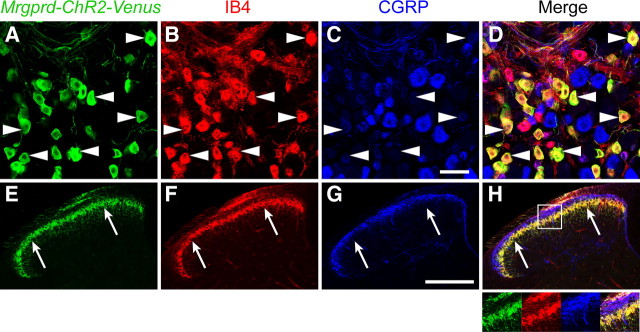
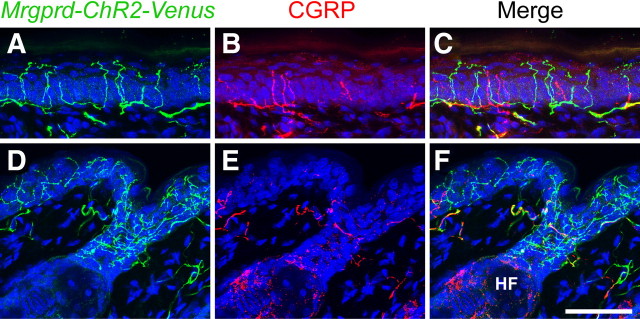
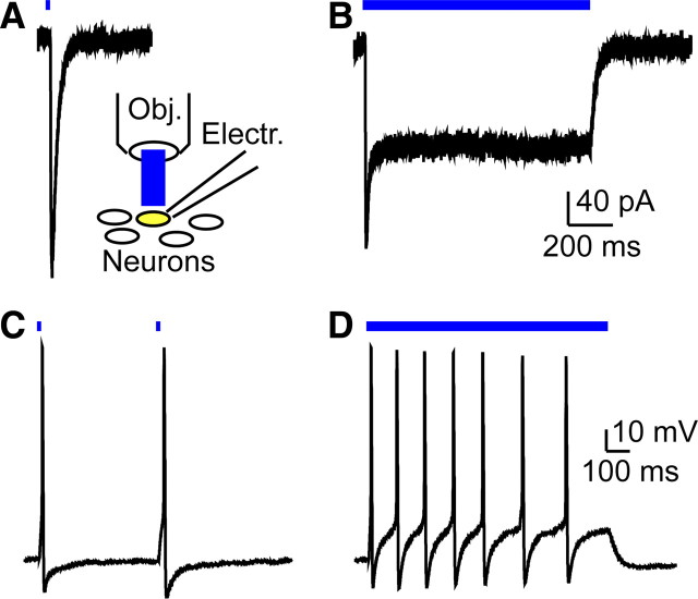
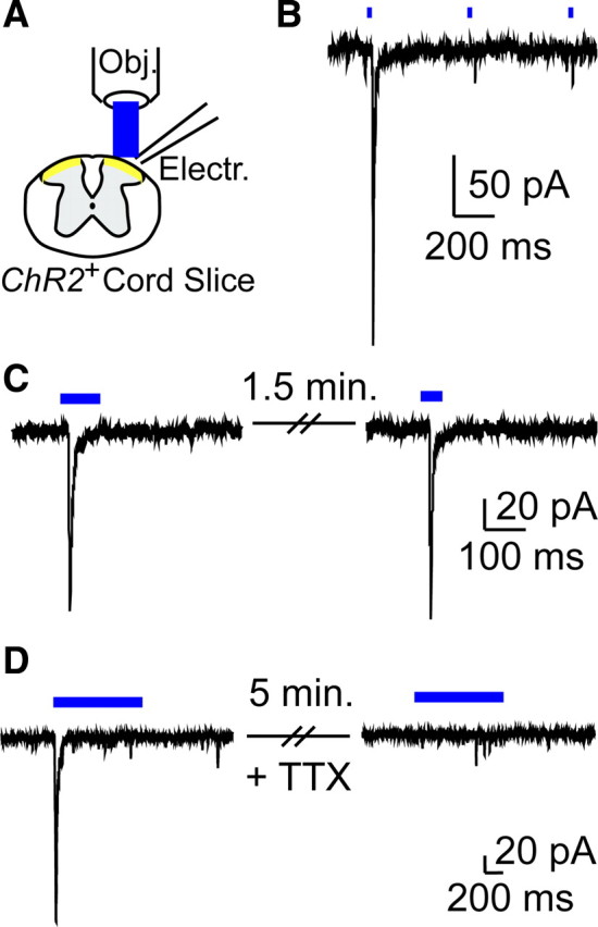
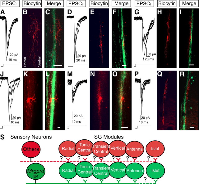
The Mas-related G-protein-coupled receptor D (Mrgprd) marks a distinct subset of sensory neurons that transmit polymodal nociceptive information from the skin epidermis to the substantia gelatinosa (SG, lamina II) of the spinal cord. Moreover, Mrgprd-expressing (Mrgprd(+)) neurons are required for the full expression of mechanical but not thermal nociception. While such anatomical and functional specificity suggests Mrgprd(+) neurons might synapse with specific postsynaptic targets in the SG, precisely how Mrgprd(+) neurons interface with spinal circuits is currently unknown. To study circuit connectivity, we genetically targeted the light-activated ion channel Channelrhodopsin-2-Venus (ChR2-Venus) to the Mrgprd locus. In these knock-in mice, ChR2-Venus was localized to nonpeptidergic Mrgprd(+) neurons and axons, while peptidergic CGRP(+) neurons were not significantly labeled. Dissociated Mrgprd(+) DRG neurons from mice expressing one or two copies of ChR2-Venus could be activated in vitro as evidenced by light-evoked currents and action potentials. In addition, illumination of Mrgprd-ChR2-Venus(+) axon terminals in spinal cord slices evoked EPSCs in half of all SG neurons. Within this subset, Mrgprd(+) neurons were monosynaptically connected to most known classes of SG neurons, including radial, tonic central, transient central, vertical, and antenna cells. This cellular diversity ruled out the possibility that Mrgprd(+) neurons innervate a dedicated class of SG neuron. Our findings set broad constraints on the types of spinal neurons that process afferent input from Mrgprd(+) polymodal nociceptors.


Figures

Figure 1.
Figure 1.
Localization of Mrgprd-ChR2-Venus to nonpeptidergic neurons and their axon terminals. A–H, L4–L6 DRG neurons (A–D) and lumbar spinal cord (E–H) from Mrgprd-ChR2-Venus+/− mice were stained with antibodies against GFP (green; anti-GFP antibodies detect Venus because Venus is virtually identical to GFP at the amino acid level), the lectin IB4 (red) and with antibodies against CGRP (blue). Examples of Mrgprd-ChR2-Venus neurons that bind IB4 are indicated (arrowheads). Arrows mark the ventral boundary of lamina II. Inset in H is magnified at the bottom right. Images were acquired by confocal microscopy. Scale bars: A–D, 50 μm; E–H, 200 μm.
Figure 2.
Figure 2.
Localization of Mrgprd-ChR2-Venus to nerve terminals in glabrous and hairy skin. A–F, Mouse glabrous skin (A–C) and hairy skin (D–F) were stained with antibodies against GFP (green) and CGRP (red). Nuclei were stained with Draq5 (blue). Images were acquired by confocal microscopy. In D–F, Mrgprd-ChR2-Venus+ endings are located in the epidermis and at the neck of a hair follicle (HF). Scale bar, 50 μm.
Figure 3.
Figure 3.
Generation of light-evoked currents and action potentials in Mrgprd-ChR2-Venus+ DRG neurons. A, Inset, Recording configuration for all panels. A, B, Voltage-clamp recordings from heterozygous Mrgprd-ChR2-Venus+ DRG neuron held at −60 mV and stimulated with blue light for 4 ms (A) or 1 s (B). C, D, Current-clamp recordings from a heterozygous Mrgprd-ChR2-Venus+ neuron stimulated with blue light at 2 Hz for a 4 ms pulse duration (C) or for 1 s (D). Electr., Electrode; Obj., objective.
Figure 4.
Figure 4.
Generation of light-evoked EPSCs in SG neurons and dependence on sodium channel activity. A, Transverse spinal cord slice recording configuration for all panels. Electr., Electrode; Obj., objective. B, Voltage-clamp recording from an SG neuron in an Mrgprd-ChR2-Venus−/− spinal cord slice, stimulated with 2 Hz pulsed light. C, Successive EPSCLs from an SG neuron in an Mrgprd-ChR2-Venus−/− mouse spinal cord slice. Delay between light presentations was 1.5 min. D, EPSCL in an SG neuron from an Mrgprd-ChR2-Venus−/− mouse was blocked by 5 min bath application of 0.5 μm TTX.
Figure 5.
Figure 5.
Light-evoked EPSCs can be generated in all known classes of SG neurons. A, D, G, J, M, P, Overlay of five successive EPSCLs from SG neurons in sagittal slices from Mrgprd-ChR2-Venus−/− mice. Delay between light presentations was 1 min. Following recording, slices were stained for biocytin (red) and Venus (green, to label Mrgprd-ChR2-Venus+ axon terminals) and then imaged by confocal microscopy. Proximity of the recorded neurons to Mrgprd-ChR2-Venus+ axon terminals is shown in the merged images. Based on physiological and morphological criteria, neurons were classified as radial (A–C), tonic central (D–F), transient central (G–I), vertical (J–L), antenna (M–O), and islet (P–R). Dorsal is to the left and rostral is toward top for all panels. Scale bar, 50 μm. S, Our data suggest that Mrgprd+ afferents innervate “SG modules” made up of most known SG cell types, with the possible exception of islet cells. Since not all SG neurons are monosynaptically connected to Mrgprd+ neurons, additional SG modules may exist for other sensory neuron subtypes. Whether these additional SG modules are made up of all known SG cell types or subsets is unknown (indicated by question marks).

References

    1. Arenkiel BR, Peca J, Davison IG, Feliciano C, Deisseroth K, Augustine GJ, Ehlers MD, Feng G. In vivo light-induced activation of neural circuitry in transgenic mice expressing channelrhodopsin-2. Neuron. 2007;54:205–218. - PMC - PubMed
    1. Bessou P, Perl ER. Response of cutaneous sensory units with unmyelinated fibers to noxious stimuli. J Neurophysiol. 1969;32:1025–1043. - PubMed
    1. Bi A, Cui J, Ma YP, Olshevskaya E, Pu M, Dizhoor AM, Pan ZH. Ectopic expression of a microbial-type rhodopsin restores visual responses in mice with photoreceptor degeneration. Neuron. 2006;50:23–33. - PMC - PubMed
    1. Boyden ES, Zhang F, Bamberg E, Nagel G, Deisseroth K. Millisecond-timescale, genetically targeted optical control of neural activity. Nat Neurosci. 2005;8:1263–1268. - PubMed
    1. Braz JM, Nassar MA, Wood JN, Basbaum AI. Parallel “pain” pathways arise from subpopulations of primary afferent nociceptor. Neuron. 2005;47:787–793. - PubMed

Publication types

MeSH terms