. 2010 Jan;298(1):C149-62.
doi: 10.1152/ajpcell.00241.2009. Epub 2009 Oct 21.

Role of TRPC1 channel in skeletal muscle function

Affiliations

Role of TRPC1 channel in skeletal muscle function

Nadège Zanou et al. Am J Physiol Cell Physiol. 2010 Jan.

Abstract

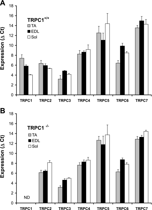
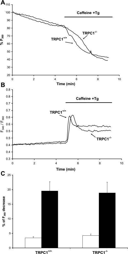
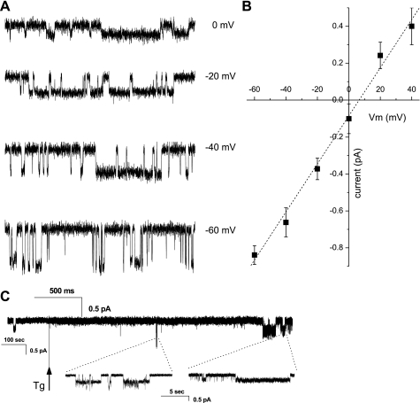
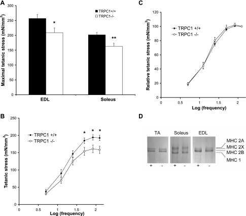
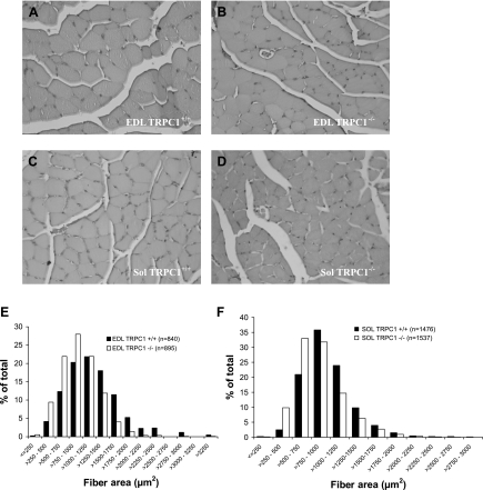
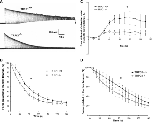
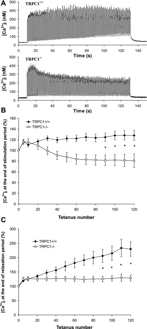
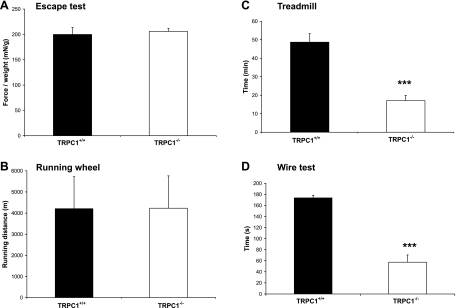
Skeletal muscle contraction is reputed not to depend on extracellular Ca2+. Indeed, stricto sensu, excitation-contraction coupling does not necessitate entry of Ca2+. However, we previously observed that, during sustained activity (repeated contractions), entry of Ca2+ is needed to maintain force production. In the present study, we evaluated the possible involvement of the canonical transient receptor potential (TRPC)1 ion channel in this entry of Ca2+ and investigated its possible role in muscle function. Patch-clamp experiments reveal the presence of a small-conductance channel (13 pS) that is completely lost in adult fibers from TRPC1(-/-) mice. The influx of Ca2+ through TRPC1 channels represents a minor part of the entry of Ca(2+) into muscle fibers at rest, and the activity of the channel is not store dependent. The lack of TRPC1 does not affect intracellular Ca2+ concentration ([Ca2+](i)) transients reached during a single isometric contraction. However, the involvement of TRPC1-related Ca2+ entry is clearly emphasized in muscle fatigue. Indeed, muscles from TRPC1(-/-) mice stimulated repeatedly progressively display lower [Ca2+](i) transients than those observed in TRPC1(+/+) fibers, and they also present an accentuated progressive loss of force. Interestingly, muscles from TRPC1(-/-) mice display a smaller fiber cross-sectional area, generate less force per cross-sectional area, and contain less myofibrillar proteins than their controls. They do not present other signs of myopathy. In agreement with in vitro experiments, TRPC1(-/-) mice present an important decrease of endurance of physical activity. We conclude that TRPC1 ion channels modulate the entry of Ca(2+) during repeated contractions and help muscles to maintain their force during sustained repeated contractions.


Figures

Fig. 1.
Fig. 1.
Quantification of canonical transient receptor potential (TRPC) channels expression in tibialis anterior (TA), extensor digitorum longus (EDL), and soleus (Sol) muscles from TRPC1+/+ and TRPC1−/− mice. A: TRPC1+/+ mice. Each cDNA was amplified in duplicate, and threshold cycle (Ct) values were averaged for each duplicate. The average Ct value for cyclophilin D was subtracted from the average Ct value for the gene of interest, giving ΔCt (n = 5 or 6). Note that gene expression is inversely proportional to ΔCt. B: TRPC1−/− mice. ΔCt values were obtained from TRPC1−/− samples (n = 4–6). ND, not detected.
Fig. 2.
Fig. 2.
Store-operated entry of Ca2+ in muscle fibers from TRPC1+/+ and TRPC1−/− mice. A and B: Ca2+ release from the stores was triggered by stimulation with 20 mM caffeine and 1 μM thapsigargin (Tg) in the presence of 1 mM Mn2+. This induced an increase of intracellular Ca2+ concentration ([Ca2+]i) (reflected by an increase of the fluorescence ratio F340/F360; B) and an increase of Ca2+ entry (reflected by an increase of the quenching rate of fura-PE3 by Mn2+; A). C: comparison of fura-PE3 quenching rates by Mn2+ at rest (gray bars) and after Ca2+ release induced by caffeine and Tg stimulation (black bars representing difference between fura-PE3 quenching rates after and before stimulation) in muscle fibers from TRPC1+/+ (n = 13) and TRPC1−/− mice (n = 14). Results are means ± SE.
Fig. 3.
Fig. 3.
Ca2+ currents in cell-attached patches. A: series of traces showing typical single-channel currents in TRPC1+/+ cells in cell-attached configuration at indicated voltages that were not observed in TRPC1−/− cells. B: current-voltage relationship of TRPC1 channel (n = 7). C: sample trace showing Ca2+ current evoked in response to sarcoplasmic reticulum (SR) emptying by thapsigargin (Tg). Patch was held at −40 mV and first basal activity was acquired, followed by the application of thapsigargin-containing solution, as indicated by vertical arrow.
Fig. 4.
Fig. 4.
Loss of TRPC1 induces a reduction of force production. A: maximal stress (force per cross-sectional area) developed during a 300-ms tetanus stimulated maximally (125 Hz). *P < 0.05 vs. TRPC1+/+; **P < 0.01 vs. TRPC1+/+ (Student's t-test, n = 9). B and C: stress vs. stimulation frequency relationship in soleus muscles. *P < 0.05 vs. TRPC1+/+ (Student's t-test, n = 9). D: SDS-PAGE analysis of myosin heavy chain (MHC) isoforms of TA, soleus, and EDL muscles from TRPC1+/+ (+) and TRPC1−/− (−) mice.
Fig. 5.
Fig. 5.
Histological data. A–D: hematoxylin and eosin-stained cross sections of EDL (A and B) and soleus (C and D) muscles from 3-mo-old TRPC1+/+ (A and C) and TRPC1−/− (B and D) mice. E and F: fiber area distribution in EDL (E) and soleus (F) muscles from TRPC1+/+ and TRPC1−/− mice. The distributions observed in TRPC1+/+ and TRPC1−/− were significantly different (P < 0.01, χ2, Pearson's test).
Fig. 6.
Fig. 6.
Involvement of TRPC1 in muscle fatigue: mechanical data. A: representative examples of force records in soleus muscles from TRPC1+/+ and TRPC1−/− mice: tetani of 500 ms every s during 2 min at 50-Hz stimulation frequency. B: quantification of the loss of force during the protocol of fatigue in soleus muscle (force measured every 10th tetanus). Results are expressed relative to the maximal force produced during 1st tetanus. Statistical analysis : *TRPC1+/+ curve (n = 10) different from TRPC1−/− curve (n = 12), P < 0.05, 1-way repeated-measures ANOVA. C: quantification of the force at the end of the relaxation period (0.5 s). Results are expressed relative to the maximal force produced during 1st tetanus. Statistical analysis: *TRPC1+/+ curve (n = 10) different from TRPC1−/− curve (n = 12), P < 0.05, 1-way repeated-measures ANOVA. D: loss of force during the protocol of fatigue in EDL muscle. Statistical analysis: *TRPC1+/+ curve (n = 7) different from TRPC1−/− curve (n = 7), P < 0.05, 1-way repeated-measures ANOVA.
Fig. 7.
Fig. 7.
Involvement of TRPC1 in muscle fatigue: [Ca2+]i transients. A: examples of [Ca2+]i transients measured in flexor digitorum brevis (FDB) fibers from TRPC1+/+ (top) and TRPC1−/− mice (bottom) submitted to the fatigue protocol described in Fig. 6. B: quantification of maximal tetanic [Ca2+]i during the protocol of fatigue ([Ca2+]i measured at 1st, 5th, and then every 10th tetanus). Results are expressed relative to maximal [Ca2+]i obtained during 1st tetanus. C: quantification of the [Ca2+]i obtained at the end of each relaxation period during the protocol of fatigue. ([Ca2+]i measured at 1st, 5th, and then every 10th tetanus). Results are expressed relative to maximal [Ca2+]i obtained during 1st tetanus. Statistical analysis: *TRPC1+/+ (n = 13) different from TRPC1−/− (n = 22), P < 0.05, 1-way repeated-measures ANOVA followed by Bonferroni multiple-comparison test.
Fig. 8.
Fig. 8.
In vivo testing. A: escape test: whole body force developed in response to a stimulus (gentle pinching of the tail). Procedure was repeated at short intervals for 2.5 min. Results are presented as means of the 5 highest peaks of force recorded, relative to body weight (mN/g) (n = 10 for each strain). B: running wheel: spontaneous running activity measured during the active part of the day (n = 11 for each strain). C: forced treadmill exercise: duration of exercise before exhaustion. ***P < 0.001 vs. TRPC1+/+ (Student's t-test, n = 10) D: wire test: mice were suspended from a horizontal metallic wire. Time until mice completely released their grasp and fell down is shown; scores of the 3 trials were averaged. ***P < 0.001 vs. TRPC1+/+ (Student's t-test, n = 10).

References

    1. Abramowitz J, Birnbaumer L. Physiology and pathophysiology of canonical transient receptor potential channels. FASEB J 23: 297–328, 2009 - PMC - PubMed
    1. Allen DG. Skeletal muscle function: role of ionic changes in fatigue, damage and disease. Clin Exp Pharmacol Physiol 31: 485–493, 2004 - PubMed
    1. Allen DG, Lamb GD, Westerblad H. Skeletal muscle fatigue: cellular mechanisms. Physiol Rev 88: 287–332, 2008 - PubMed
    1. Allen DG, Lannergren J, Westerblad H. Muscle cell function during prolonged activity: cellular mechanisms of fatigue. Exp Physiol 80: 497–527, 1995 - PubMed
    1. Allen DG, Westerblad H. Role of phosphate and calcium stores in muscle fatigue. J Physiol 536: 657–665, 2001 - PMC - PubMed

Publication types

MeSH terms

LinkOut - more resources