. 2009 Nov 3;106(44):18527-32.
doi: 10.1073/pnas.0906132106. Epub 2009 Oct 21.

Insights into the reactivation of cobalamin-dependent methionine synthase

Affiliations

Insights into the reactivation of cobalamin-dependent methionine synthase

Markos Koutmos et al. Proc Natl Acad Sci U S A. .

Abstract

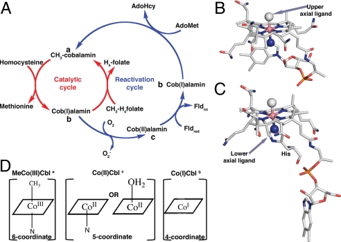
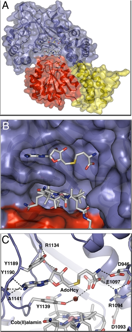
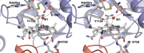
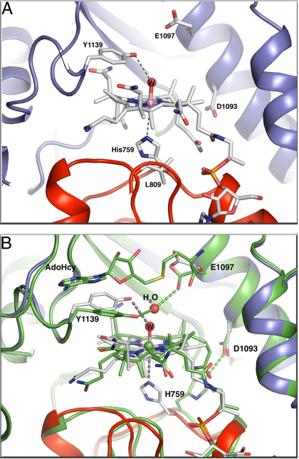
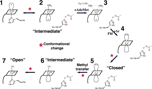
Cobalamin-dependent methionine synthase (MetH) is a modular protein that catalyzes the transfer of a methyl group from methyltetrahydrofolate to homocysteine to produce methionine and tetrahydrofolate. The cobalamin cofactor, which serves as both acceptor and donor of the methyl group, is oxidized once every approximately 2,000 catalytic cycles and must be reactivated by the uptake of an electron from reduced flavodoxin and a methyl group from S-adenosyl-L-methionine (AdoMet). Previous structures of a C-terminal fragment of MetH (MetH(CT)) revealed a reactivation conformation that juxtaposes the cobalamin- and AdoMet-binding domains. Here we describe 2 structures of a disulfide stabilized MetH(CT) ((s-s)MetH(CT)) that offer further insight into the reactivation of MetH. The structure of (s-s)MetH(CT) with cob(II)alamin and S-adenosyl-L-homocysteine represents the enzyme in the reactivation step preceding electron transfer from flavodoxin. The structure supports earlier suggestions that the enzyme acts to lower the reduction potential of the Co(II)/Co(I) couple by elongating the bond between the cobalt and its upper axial water ligand, effectively making the cobalt 4-coordinate, and illuminates the role of Tyr-1139 in the stabilization of this 4-coordinate state. The structure of (s-s)MetH(CT) with aquocobalamin may represent a transient state at the end of reactivation as the newly remethylated 5-coordinate methylcobalamin returns to the 6-coordinate state, triggering the rearrangement to a catalytic conformation.


Conflict of interest statement

The authors declare no conflict of interest.

Figures

Fig. 1.
Fig. 1.
(A) Reaction cycle of MetH. The primary catalytic cycle is shown in red whereas the secondary reactivation cycle is shown in blue. (B) MeCo(III)Cbl in the free state with a N atom of the dimethylbenzimidazole group coordinating Co in the lower axial position. A methyl ligand occupies the upper axial position. (C) MetH-bound MeCo(III)Cbl with H759 as the lower axial ligand (13). (D) Schematic drawings of the preferred coordination numbers and geometries for the Cbl oxidation states that are relevant to MetH function. The superscripts a, b, and c in part A indicate the locations of these forms of the cofactor in the catalytic and reactivation cycles. The corrin ring of the Cbl is depicted as a square for simplicity.
Fig. 2.
Fig. 2.
(A) S-SMetHCT:Co(II)Cbl:AdoHcy complex ribbon diagram and space-filling model with the AdoMet-binding module in blue, the Cbl-binding domain in red, and the Cap domain (the N-terminal domain of the Cbl-binding module) in yellow. (B) Surface rendering of the Cbl binding pocket below the AdoHcy binding pocket with subunits colored as in A. AdoHcy and Co(II)Cbl are shown as sticks with white C, red O, blue N, yellow S, and pink Co. (C) AdoHcy binding site. The AdoMet-binding domain is shown as a blue ribbon, with AdoHcy, Co(II)Cbl and amino acid side chains in the binding site in stick form and a water molecule as a red sphere. Hydrogen bonds are shown as dashed lines; colors as in B.
Fig. 3.
Fig. 3.
Stabilization of 4-coordinate Co(II)Cbl. The protein environment promotes transition of the metal from a 5-coordinate to a 4-coordinate state through hydrogen bonds to E1097 and Y1139 that move the water away from the Co. This cross-eyed stereoview of the active site of the S-SMetHCT:Co(II)Cbl:AdoHcy complex shows the important interactions that enable the Co–OH2 bond elongation. The water molecule formally bound to Cbl in the absence of AdoHcy is designated as W1. The distances indicated with gray dotted lines are: Y1139-OH–Co(II), 3.9 Å; W1–Co(II), 3.6 Å; Y1139-OH–W1, 3.3 Å; E1097-O–W1, 3.1 Å.
Fig. 4.
Fig. 4.
Spectrophotometric determination of the Co(II)Cbl/Co(I)Cbl midpoint potential of S-SMetHCT (A) and Y1139F S-SMetHCT (B). The enzyme was first photoreduced with 5-deazaflavin-3-sulfonate in the presence of the indicator dye methyl viologen to generate a Co(I)Cbl-bound species (as evidenced by the decrease in absorbance at 468 nm), and spectra were then recorded to monitor the increase in absorbance at 468 nm associated with the spontaneous reoxidation to the Co(II)Cbl-bound form. From a Nernst plot the midpoint potentials were estimated to be −490 mV and −540 mV for S-SMetHCT and Y1139F S-SMetHCT (C and D).
Fig. 5.
Fig. 5.
(A) Environment of the AquoCo(III)Cbl active site in S-SMetHCT:AquoCo(III)Cbl. The distances indicated with gray dotted lines are: Y1139-OH–W, 3.3 Å; H759-Nε2–Co(III), 3.5 Å. The Co–H2O bond distance is 2.1 Å. (B) Superposition of the active sites of the S-SMetHCT:Co(II)Cbl:AdoHcy complex (green protein and green C atoms) and the S-SMetHCT:AquoCo(III)Cbl structure (color scheme as in Fig. 1). Superpositions are based on residues from the ß-strands of the AdoMet domain. The position of the AdoMet-binding domain and the corrin ring is basically unchanged between the 2 structures. The cobalamin-binding domain, however, moves as a rigid body with respect to the AdoMet-binding domain and the corrin ring.
Fig. 6.
Fig. 6.
Schematic diagram of the changes in cobalt coordination oxidation state and environment during the reactivation cycle. The corrin ring of the Cbl is depicted as a rhomboid and the dimethylbenzimidazole as a line at a corner of the square. (1) Co(II)Cbl is 5-coordinate, with H759 occupying the lower axial position. (2) When Fld binds to the full-length MetH, or in S-SMetHCT, a different 5-coordinate Co(II)Cbl species is seen in which the lower axial His-ligand is replaced with an upper axial water ligand (19, 20). This step is associated with a conformational change from a catalytic conformation in which the cobalamin-binding module is juxtaposed with a Hcy-binding or CH3-H4folate-binding module to the reactivation conformation where the cobalamin-binding module is juxtaposed to the AdoMet-binding module. (3) AdoMet/AdoHcy binding leads to water-Co(II) bond elongation, represented by the AdoHcy-bound Co(II)Cbl S-SMetHCT structure presented here. Fld association and electron transfer lead to the formation of an “intermediate” 4-coordinate Co(I)Cbl species (4) and conformational changes that bring the Me group closer to the Co (5) and allow the subsequent methyl transfer in this “closed” conformation. Methyl transfer and formation of a 5-coordinate MeCo(III)Cbl species allows the domains to return to the “intermediate” state (6). Finally, domain motion is required for the His-759 to again coordinate the Co atom to form the 6-coordinate MeCo(III)Cbl species (7). This “open” conformation is represented by the AquoCo(III)Cbl structure presented here and is the last step before the Cob domain switches from the reactivation cycle back to the catalytic cycle where it can interact with substrate-binding domains and support catalysis.

References

    1. Goulding CW, Postigo D, Matthews RG. Cobalamin-dependent methionine synthase is a modular protein with distinct regions for binding homocysteine, methyltetrahydrofolate, cobalamin, and adenosylmethionine. Biochemistry. 1997;36:8082–8091. - PubMed
    1. Matthews RG, Koutmos M, Datta S. Cobalamin-dependent and cobamide-dependent methyltransferases. Curr Opin Struct Biol. 2008;18:658–666. - PMC - PubMed
    1. Drummond JT, Huang S, Blumenthal RM, Matthews RG. Assignment of enzymatic function to specific protein regions of cobalamin-dependent methionine synthase from Escherichia coli. Biochemistry. 1993;32:9290–9295. - PubMed
    1. Fujii K, Galivan JH, Huennekens FM. Activation of methionine synthase: Further characterization of flavoprotein system. Arch Biochem Biophys. 1977;178:662–670. - PubMed
    1. Taylor RT, Weissbach H. N5-methyltetrahydrofolate-homocysteine transmethylase: Partial purification and properties. J Biol Chem. 1967;242:1502–1508. - PubMed

Publication types

MeSH terms

LinkOut - more resources