

Cerenkov radiation imaging as a method for quantitative measurements of beta particles in a microfluidic chip
- PMID: 19847018
- PMCID: PMC2794558
- DOI: 10.1088/0031-9155/54/22/001
Cerenkov radiation imaging as a method for quantitative measurements of beta particles in a microfluidic chip
Abstract
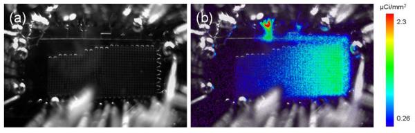
It has been observed that microfluidic chips used for synthesizing (18)F-labeled compounds demonstrate visible light emission without nearby scintillators or fluorescent materials. The origin of the light was investigated and found to be consistent with the emission characteristics from Cerenkov radiation. Since (18)F decays through the emission of high-energy positrons, the energy threshold for beta particles, i.e. electrons or positrons, to generate Cerenkov radiation was calculated for water and polydimethylsiloxane (PDMS), the most commonly used polymer-based material for microfluidic chips. Beta particles emitted from (18)F have a continuous energy spectrum, with a maximum energy that exceeds this energy threshold for both water and PDMS. In addition, the spectral characteristics of the emitted light from (18)F in distilled water were also measured, yielding a broad distribution from 300 nm to 700 nm, with higher intensity at shorter wavelengths. A photograph of the (18)F solution showed a bluish-white light emitted from the solution, further suggesting Cerenkov radiation. In this study, the feasibility of using this Cerenkov light emission as a method for quantitative measurements of the radioactivity within the microfluidic chip in situ was evaluated. A detector previously developed for imaging microfluidic platforms was used. The detector consisted of a charge-coupled device (CCD) optically coupled to a lens. The system spatial resolution, minimum detectable activity and dynamic range were evaluated. In addition, the calibration of a Cerenkov signal versus activity concentration in the microfluidic chip was determined. This novel method of Cerenkov radiation measurements will provide researchers with a simple yet robust quantitative imaging tool for microfluidic applications utilizing beta particles.
Figures


References
-
- Andersson H, van den Berg A. Microfluidic devices for cellomics: a review. Sensors and Actuators B: Chemical. 2003;92:315–325.
-
- Bogalhas F, Menard L, Bonzom S, Palfi S, Siebert R, Duval M, Lefebvre F, Pinot L, Pitre S, Charon Y. Physical performance of an intraoperative beta probe dedicated to glioma radioguided surgery. IEEE Transactions on Nuclear Science. 2008;55(no 3):833–841.
-
- Breskin A. Advances in gas avalanche radiation detectors for biomedical applications. Nucl. Instr. Meth. 2000 January;A454:26–39. 2000.
-
- Cabello J, Bailey A, Kitchen I, Prydderch M, Clark A, Turchetta R, Wells K. Digital autoradiography using room temperature CCD and CMOS imaging technology. Phys. Med. Biol. 2007;52:4993–5011. - PubMed
-
- Cherry SR, Sorenson JA, Phelps ME. Physics in Nuclear Medicine. Saunders; Philadelphia: 2003.
Publication types
MeSH terms
Substances
Grants and funding
LinkOut - more resources
Full Text Sources
Other Literature Sources