. 2010 Mar;4(1):51-62.
doi: 10.1007/s12079-009-0073-3. Epub 2009 Oct 22.

Endothelin-1 as a neuropeptide: neurotransmitter or neurovascular effects?

Endothelin-1 as a neuropeptide: neurotransmitter or neurovascular effects?

Michael R Dashwood et al. J Cell Commun Signal. 2010 Mar.

Abstract

Endothelin-1 (ET-1) is an endothelium-derived peptide that also possesses potent mitogenic activity. There is also a suggestion the ET-1 is a neuropeptide, based mainly on its histological identification in both the central and peripheral nervous system in a number of species, including man. A neuropeptide role for ET-1 is supported by studies showing a variety of effects caused following its administration into different regions of the brain and by application to peripheral nerves. In addition there are studies proposing that ET-1 is implicated in a number of neural circuits where its transmitter affects range from a role in pain and temperature control to its action on the hypothalamo-neurosecretory system. While the effect of ET-1 on nerve tissue is beyond doubt, its action on nerve blood flow is often ignored. Here, we review data generated in a number of species and using a variety of experimental models. Studies range from those showing the distribution of ET-1 and its receptors in nerve tissue to those describing numerous neurally-mediated effects of ET-1.


Figures
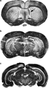
Fig. 1
Fig. 1
Representative frontal sections of rat brain. Frontal sections of rat brain (Sudan black staining) at three different levels from rostral a to caudal c showing: in a the lateral ventricles (arrow), in b third ventricle (arrow) and thalamus-hypothalamus with supraoptic nucleus (asterisk), and in c showing aqueduct (arrow) and periaquedutcal gray area (asterisk). ET-1 adiministration to such regions has cardiovascular (D’Amico et al. ; McAuley et al. ; Macrae et al. , ; Macrae et al. 1993) and temperature-regulating effects (Fabricio et al. 2005)
Fig. 2
Fig. 2
Cerebral vessels. Left panel. Magnification angiography of normal rat brain using barium sulphate contrast medium. There is an abundant vascular supply with many penetrating microvessels in the cortex shown in the right panel following intra-arterial injection of India ink (black regions of a haemotoxylin and eosin stains section). Scale bar = 5 mm for the left panel and 50 µm for the right panel. Modified from Hekmatpanah Surg Neurol 2007;67:564-71
Fig. 3
Fig. 3
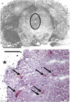
ET-1 action on the periaqueductal gray area of the rat. Top panel. ETA receptors ([125I-PD 151252 binding) in the rat brain identified by in vitro autoradiography (from D’Amico et al. 1996). Periaqueductal gray area (PAG) is outlined. Lower panel. Cerebral vessels of the PAG identified by immunohistochemistry (arrows indicate CD31 staining of vessel endothelium, red). (Dashwood unpublished) *indicates the aqueduct. Scale bars = 2.5 mm for the top and 50 µm for the lower panels
Fig. 4
Fig. 4
Neuropil of supraoptic nucleus of the hypothalamus double-labelled for nNOS (immunoprecipitate - long arrows) and ET-1 (immunogold-silver grains – short arrows). a A fragment of a double-labelled dendrite (likely to be a primary or varicose dendrite from a bipolar magnocellular neurone) shows abundance of neurosecretory granules (nsg), of which some are nNOS-positive (long arrows); short arrows point to immunoreactivity for ET-1. Note unlabelled axon profile (Ax) making synapse on the double-labelled dendrite. b A double-labelled axon terminal (At) making asymmetric synapse with unlabelled dendrite spine (ds). The axon terminal contains numerous small agranular synaptic vesicles, membrane of which is labelled with nNOS immunoprecipitate; co-localised immunogold-silver grains of ET-1 labelling (arrows) are also seen. Note that an adjacent axon profile (Ax) containing small agranular synaptic vesicles is unlabelled; m-mitochondria. Bars: 0.5 μm. Affinity-purified rabbit polyclonal nNOS antibody (SC-1025, Santa Cruz, USA), which does not cross react with NOS2 and NOS3 was used at 0.8μg/ml in the preembedding ExtrAvidin immunocytochemical method. The rabbit polyclonal ET-1 antibody to human/porcine ET-1 (Sigma, Poole, UK), which does not cross-react with big ET but may with ET-2 and ET-3 was used at 1:1,000 in the preembedding immunogold-silver labelling method as the second immunolabelling. Images A and B are modified from Mukherjee and Loesch, Histochem J 2002, 34:181-187 [Kluwer Academic Publishers], which is kindly acknowledged
Fig. 5
Fig. 5
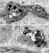
Perivascular nerve/axon varicosities in basilar artery of a normal (A) and hypertensive (B) rats immunolabelled for ET-1. a Note granular (gv) and agranular (av) vesicles; core of granular vesicles is intensely labelled for ET-1. In B note damaged axon varicosity with clustered agranular vesicles (av), vacuoles with dense material (v), and ‘empty’ areas of axoplasm (asterisk). Adjacent ET-1-negative varicosity (Ax) is of normal appearance with evenly distributed vesicles. Bars: 0.2 µm. Rabbit polyclonal ET-1 antibodies to human/porcine ET-1 (a from CRB, Cambridge, UK; b from Sigma, Poole, UK) was used at 1:1,000 in the preembedding PAP method; antibodies do not cross-react with big ET but may with ET-2 and ET-3. A is modified from Loesch et al. Neuroreport 1998, 9:3903-3906 [Lippincott Williams & Wilkins] and B is from Milner et al. J Vasc Res 2000; 37:39-49 [S Karger AG, Basel/Medical and Science publishers], which is kindly acknowledged
Fig. 6
Fig. 6
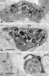
Capybara basilar artery perivascular nerves labelled (black precipitate) for ETA (A) and ETB (B) receptors. In a and b note axons (Ax) and Schwann cell profiles (Sch) displaying immunoreactivity (arrows) for ETA and ETB receptors, respectively; some labelling is seen in the granular vesicles. In C note ETB localisation in the Schwann cells only, while associated axon varicosities is negative for ETB receptors. Bars: 0.5 µm. Rabbit polyclonal antibodies ETA and ETB (Alomone Labs, Jerusalem, Israel) were used at 1:400 in pre-embedding ExtrAvidin method. The ETA receptor antibody (AER-001) recognises intracellular (C-terminus) epitope corresponding to amino acid residues 413-426 of rat ETA peptide (Accession P26684), while the ETB receptor antibody (AER-002) recognises intracellular (i3 loop) epitope corresponding to residues 298-314 of rat ETB peptide (Accession P21451). Both antibodies were affinity purified on immobilized antigens. (A-C are modified from Loesch et al., J Mol Histol 2005; 36: 25-34 [Kluwer and Springer Science and Business Media], which is kindly acknowledged)
Fig. 7
Fig. 7
Endothelin and its receptors on human sural nerve. a Location of human sural nerve: (From Aktan Ikiz et al. 2005) sn:sural nerve, lm:lateral malleolus, idcn: intermediate dorsal cutaneous nerve. b Distribution of ET-1, ETA and ETB receptors on human sural nerve sections revealed by in vitro autoradiography using radiation-sensitive film (low resolution). c Microscopic localisation of ET-1 on a section of human sural nerve. Left panel is a dark-field illumination, ‘high resolution’ autoradiograph of a section incubated in 125I-labelled ET-1 and detected using nuclear emulsion where white grains show receptor binding sites. Right panel is the haemotoxylin and eosin stained tissue. There is strong binding to the epineurial vessels (BVS), perineurium (arrowheads) and vasa nervorum (small cell clusters within the nerve fascicles surrounded by the perineurium). Scale bars = 2 mm for autoradiographs in B and 50 µm for C (B and C modified from Dashwood and Thomas 1997)

References

    1. Ahn YM, Gajdusek C, London S, Moon CT, Oh CW, Mayberg MR. Sustained arterial narrowing after prolonged exposure to perivascular endothelin. Neurosurgery. 2002;50:843–848. doi: 10.1097/00006123-200204000-00029. - DOI - PubMed
    1. Aktan Ikiz ZA, Ucerler H, Bilge O. The anatomic features of the sural nerve with an emphasis on its clinical importance. Foot Ankle Int. 2005;26:560–567. - PubMed
    1. Arai H, Hori S, Aramori I, Ohkubo H, Nakanishi S. (1990) Cloning and expression of a cDNA encoding and endothelin receptor. Nature. 1990;348:730–732. doi: 10.1038/348730a0. - DOI - PubMed
    1. Barti-Mattera LN, Harwalkar S, Huges B, Wilkins PL, Almhanna K. Proliferative and morphological effects of endothelins in Schwann cells: roles of p38 mitogen-activated protein kinase and Ca2+-independenta phospholipase A2. J Neurochem. 2001;79:1136–1148. doi: 10.1046/j.1471-4159.2001.00642.x. - DOI - PubMed
    1. Buller KM, Khanna S, Sibbald JR, Day TA. Central noradrenergic neurons signal via ATP to elicit vasopressin responses to haemorrhage. Neuroscience. 1996;73:637–642. doi: 10.1016/0306-4522(96)00156-X. - DOI - PubMed